


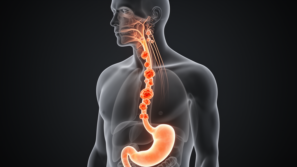
A 51-year-old man in China did everything right. When screening endoscopy revealed an early tumor in the middle of his esophagus, surgeons removed it endoscopically, then followed guidelines to the letter by performing a radical esophagectomy with extensive lymph node dissection just two weeks later. Every lymph node removed during the operation came back negative for cancer. Yet nine months after surgery, a massive tumor erupted in the lymph nodes of his neck, wrapping around his carotid artery and invading a major vein. A new case report published in Frontiers in Oncology dissects this alarming clinical course and argues that the field’s standard risk-assessment playbook contains a dangerous blind spot.
… [continue with 8-10 paragraphs total, 1400-1600 words]
The case hinges on a pathological classification system that many clinicians outside of high-volume esophageal centers may find unfamiliar. The Japanese Esophageal Society’s INF grading divides tumors into three categories based on the character of their advancing edge: INFa describes an expansile, sharply demarcated growth pattern, INFc describes a diffusely infiltrative front that blends imperceptibly into surrounding tissue, and INFb sits between the two, combining features of both. This is not merely a descriptive exercise. Studies from Japanese surgical cohorts have demonstrated that the infiltrative patterns carry independent prognostic weight, correlating with higher rates of lymph node metastasis, lymphatic permeation, and poorer survival even after adjustment for depth of invasion and lymphovascular involvement. The biological rationale is intuitive: a tumor front that disperses as individual cells or small nests rather than pushing as a cohesive mass has far greater opportunity to encounter and invade thin-walled lymphatic channels, seeding nodes at a distance before the primary lesion itself has grown substantially.
What makes the INFb category particularly treacherous in practice is its intermediate position. Guidelines and risk calculators built around the 200-micrometer submucosal invasion threshold and lymphovascular status were largely validated in populations where INF status was either recorded inconsistently or not weighted heavily in treatment algorithms. A pathologist reading a slide of an INFb tumor sees a front that is neither cleanly pushing nor obviously scattered, and interobserver agreement for this middle category is known to be weaker than for the extremes. The result is a systematic underestimation of risk: tumors classified as INFb are often treated clinically as if they behave like INFa lesions, when their metastatic behavior more closely resembles, or in some series approaches, that of INFc tumors. The authors of this case report argue that this ambiguity is precisely where patients fall through the cracks.
The anatomy of the middle thoracic esophagus compounds the problem. During embryonic development, the cervical esophagus and the upper thoracic segment share a common lymphatic watershed, and the mid-esophagus sits at a junctional zone where lymphatic drainage can flow bidirectionally—upward toward the supraclavicular and cervical basins or downward toward the mediastinal and celiac stations. Japanese anatomical studies using dye injection and radioisotope tracing have shown that lymphatic channels from the middle third of the thoracic esophagus can reach the cervical paraesophageal nodes, designated 101 in the JES numbering system, without traversing any intermediate station that a surgeon would typically harvest. This so-called skip metastasis phenomenon is well documented in esophageal cancer generally, with skip lesions reported in a meaningful minority of node-positive patients, but it becomes clinically consequential when the primary tumor carries an infiltrative phenotype that favors lymphatic dissemination early in its natural history.
The two-field lymphadenectomy performed in this patient—encompassing the mediastinal and abdominal nodal stations—is the standard operation for thoracic esophageal cancer in most East Asian centers and is increasingly adopted elsewhere. It deliberately excludes the cervical field, which is reserved for three-field dissection, a more extensive operation associated with higher morbidity that some Japanese centers advocate for tumors with extensive nodal disease or specific risk profiles. The decision of whether to extend dissection into the neck is typically driven by preoperative imaging and the perceived nodal risk of the tumor. In this case, the tiny left 101L node visible on preoperative CT was the one signal that something might be amiss in the cervical basin, and it was discounted precisely because the patient’s tumor appeared to have every reassuring feature: superficial depth, negative lymphovascular invasion, and negative margins. The case report suggests that this reasoning inverted the correct logic. A suspicious node on imaging, however small, in a drainage basin connected to the tumor’s location should prompt further investigation—ideally metabolic imaging with fluorodeoxyglucose positron emission tomography combined with computed tomography, and in select cases tissue sampling—rather than dismissal based on the primary tumor’s apparent low-risk profile.
The temporal pattern of the recurrence also deserves scrutiny. Surveillance CT at one month and again at five months after surgery showed no abnormality, yet by nine months the cervical mass had reached six centimeters in its greatest dimension and had invaded the internal jugular vein as a tumor thrombus. This trajectory implies a doubling behavior that is extraordinarily aggressive for a lesion that was undetectable five months earlier, or alternatively a lesion that was present but radiologically occult at the five-month scan—plausible for a small node nestled among the vessels and fat of the supraclavicular fossa on conventional contrast-enhanced CT. Either interpretation carries the same lesson: the interval between surveillance scans in the first postoperative year, when recurrence risk is highest, may be too long for patients whose tumors carry unweighted risk factors, and thin-slice imaging of the neck should be considered part of the standard surveillance protocol rather than an optional addition.
Tumor thrombus within the internal jugular vein is a rare but recognized complication of advanced cervical nodal disease, and it fundamentally alters the therapeutic calculus. Once tumor has breached the vein wall and propagated as a thrombus, the possibility of hematogenous dissemination rises sharply, and complete surgical resection of the nodal mass with vascular reconstruction becomes both technically formidable and oncologically questionable. This is why the treating team pivoted to concurrent chemoradiotherapy, delivering fifty gray in twenty-five fractions to the supraclavicular drainage region, with the nodal mass showing a partial response after four cycles of systemic therapy. The planned continuation with chemotherapy followed by sequential immunotherapy reflects the current paradigm in esophageal cancer, where immune checkpoint inhibitors targeting the programmed cell death pathway have demonstrated survival benefits in advanced and recurrent disease and are increasingly woven into multimodal treatment sequences.
For the broader clinical community, the case raises the question of how INF classification should be operationalized outside of Japan, where it is part of the standard synoptic pathology report but remains inconsistently incorporated into Western practice. The College of American Pathologists and equivalent bodies in Europe do not currently mandate INF reporting for esophageal specimens, meaning that many resection specimens worldwide are never formally graded for infiltration pattern. If the accumulating evidence that INF status independently predicts nodal metastasis is confirmed in larger Western cohorts, the simplest intervention would be to require INF grading in all esophageal endoscopic resection and esophagectomy specimens, giving treating teams a data point that currently goes unrecorded. The cost of this change is negligible; the pathology already exists on the slide, and the grading requires only a trained eye and a standardized reporting template.
The second practical implication concerns the management of indeterminate cervical nodes seen on preoperative cross-sectional imaging in patients with superficial esophageal cancer. The prevalence of such findings is not trivial, and the instinct to dismiss them in the setting of an otherwise low-risk tumor is understandable given the morbidity of extended lymphadenectomy and the desire to avoid over-treatment. But this case demonstrates that the perceived risk hierarchy can fail catastrophically when an unmeasured variable—infiltrative growth pattern—shifts the true probability of cervical dissemination upward. A low threshold for obtaining fluorodeoxyglucose PET-CT in this scenario, as current staging guidelines already suggest, would have revealed metabolic activity in the tiny 101L node in this patient and could have prompted either preoperative tissue diagnosis or an extended dissection that included the cervical field. The authors stop short of recommending three-field dissection for all INFb tumors, which would expose many patients to unnecessary morbidity, but they make a persuasive argument that the combination of mid-esophageal location, INFb pattern, and any cervical imaging abnormality should trigger individualized escalation rather than protocol-driven reassurance.
Finally, the case is a reminder that case reports, though often undervalued in the evidence hierarchy, serve an essential function in identifying failure modes that large trials are statistically powered to average away. A single patient who followed every guideline and still developed an explosive, vessel-invading recurrence within nine months is precisely the kind of signal that should prompt the field to re-examine its assumptions. The convergence of an intermediate infiltrative phenotype, a mid-esophageal primary with bidirectional lymphatic access to the neck, and a dismissed imaging finding created a cascade in which each individually reasonable decision compounded into a devastating outcome. Rectifying this will require not a single dramatic change but a series of modest ones: mandatory INF reporting, disciplined adherence to metabolic imaging guidelines when cervical nodes appear on preoperative scans, individualized decisions about the extent of lymphadenectomy, and intensified thin-slice surveillance of both the neck and chest during the vulnerable first postoperative year.
Subject of Research: Cervical lymph node recurrence after surgery for superficial esophageal squamous cell carcinoma with an infiltrative growth pattern
Article Title: Explosive cervical lymph node recurrence following two-field lymphadenectomy in pT1b (SM2) middle esophageal squamous cell carcinoma with INFb pattern and absent lymphovascular invasion: a case report
Article References: Explosive cervical lymph node recurrence following two-field lymphadenectomy in pT1b (SM2) middle esophageal squamous cell carcinoma with INFb pattern and absent lymphovascular invasion: a case report. (n.d.). https://doi.org/10.3389/fonc.2026.1976165
Image Credits: AI Generated
DOI: 10.3389/fonc.2026.1976165
Keywords: esophageal cancer, squamous cell carcinoma, lymph node metastasis, endoscopic submucosal dissection, lymphadenectomy, tumor infiltration pattern, PET-CT, case report, cancer recurrence, surgical oncology, lymphovascular invasion, immunotherapy
Cite Scienmag News
Nathaniel Bowman. (October 16, 2026). How a ‘Low-Risk’ Esophageal Cancer Defied the Rules and Struck the Neck Within Months. Scienmag. https://scienmag.com/how-a-low-risk-esophageal-cancer-defied-the-rules-and-struck-the-neck-within-months/
Nathaniel Bowman. "How a ‘Low-Risk’ Esophageal Cancer Defied the Rules and Struck the Neck Within Months." Scienmag, 16 October 2026, https://scienmag.com/how-a-low-risk-esophageal-cancer-defied-the-rules-and-struck-the-neck-within-months/. Accessed 16 October 2026.
Nathaniel Bowman. "How a ‘Low-Risk’ Esophageal Cancer Defied the Rules and Struck the Neck Within Months." Scienmag. October 16, 2026. https://scienmag.com/how-a-low-risk-esophageal-cancer-defied-the-rules-and-struck-the-neck-within-months/


