


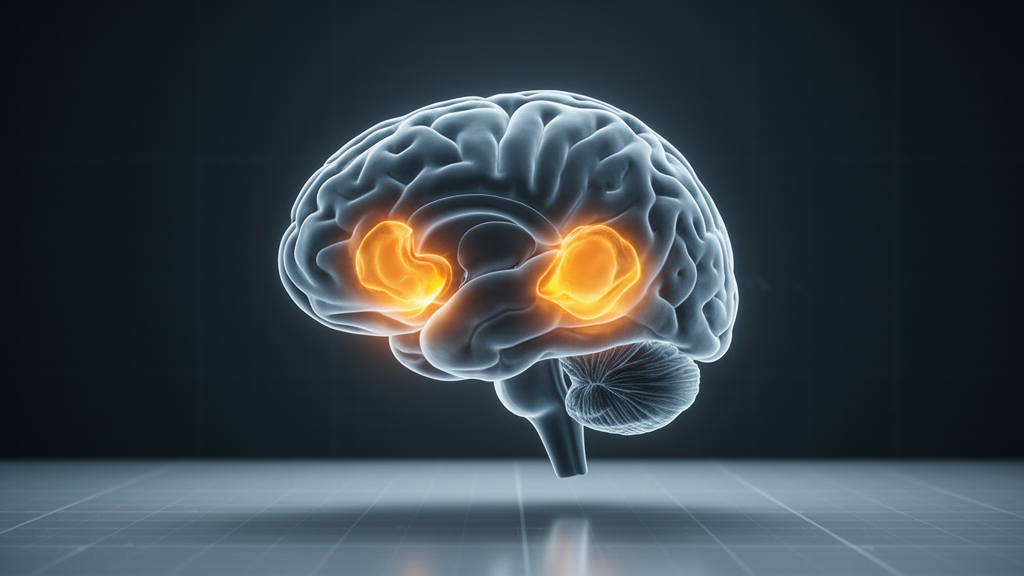
A 58-year-old man walked into a German hospital with memory trouble and seizures that seemed to come from nowhere. His initial brain MRI had been reported as normal, his neurological examination was unremarkable, and standard antibody tests came back negative. Yet when researchers at Goethe University Frankfurt applied advanced quantitative imaging techniques to his case, they uncovered evidence of brain inflammation that conventional scans had missed entirely — and their findings, published in Frontiers in Neurology, suggest that routine imaging may systematically underestimate the true extent of a rare autoimmune brain disease called limbic encephalitis.
Limbic encephalitis is an inflammatory condition that targets the limbic system, a network of deep brain structures including the hippocampus and amygdala that are critical for memory and emotion. The disease typically produces subacute cognitive decline, personality changes, and seizures, and it is often triggered by the immune system mistakenly attacking brain tissue. In systemic sclerosis — the autoimmune connective tissue disease the patient had been living with since 2021 — neurological complications are usually attributed to blood vessel damage rather than direct inflammation of brain parenchyma, making this case particularly unusual and diagnostically challenging.
When the patient was admitted in January 2026 with progressive confusion, memory loss, and episodes of absent awareness, his clinical team faced a diagnostic puzzle. Prolonged video-electroencephalography monitoring captured abnormal electrical activity from the right temporal lobe, pointing toward a seizure focus in the brain’s temporal region. But his routine brain MRI showed only subtle, unilateral changes in the right mesial temporal structures — findings that fell short of the bilateral involvement required by current diagnostic criteria for definite autoimmune limbic encephalitis. Without those criteria met, the team turned to quantitative imaging methods that go beyond what the naked eye can see on standard scans.
The researchers deployed two complementary advanced techniques. The first, T2 relaxometry, measures how quickly protons in brain tissue return to their equilibrium state after being excited by radiofrequency pulses in the MRI scanner — a property that changes when tissue undergoes inflammation, edema, or cell damage. The second, proton magnetic resonance spectroscopy, measures the concentrations of specific brain metabolites such as N-acetylaspartate, a marker of neuronal health and density. Together, these methods provide objective, numbers-based measurements of tissue integrity rather than relying on visual interpretation alone.
The results were striking. While conventional MRI showed abnormalities confined to the right mesial temporal region, T2 relaxometry revealed significantly prolonged relaxation times bilaterally — meaning both the left and right mesial temporal structures showed evidence of tissue alteration, not just the right side. The spectroscopy data reinforced this picture: N-acetylaspartate levels were reduced on both sides, with a more pronounced decrease on the right. This pattern of bilateral, right-predominant involvement aligned with what neuropathological studies of limbic encephalitis have long suggested — that the inflammatory process often affects both temporal lobes even when only one appears abnormal on standard imaging.
The quantitative findings proved decisive for the patient’s clinical course. Based on the combined evidence from relaxometry, spectroscopy, electroencephalography, and the clinical picture, the team diagnosed antibody-negative probable autoimmune limbic encephalitis in the setting of systemic sclerosis. Treatment with the immunosuppressant cyclophosphamide — a chemotherapy agent that dampens immune system activity — was initiated alongside antiseizure medications. The patient responded well: his seizures came under control, his cognitive symptoms improved, and follow-up imaging showed stabilization of the previously identified abnormalities without progression.
The case carries broader implications for how suspected limbic encephalitis is diagnosed and classified. Current diagnostic criteria, established by international consensus panels, require bilateral mesial temporal abnormalities on conventional MRI for a diagnosis of definite autoimmune limbic encephalitis. Patients with unilateral findings are instead categorized as having possible autoimmune encephalitis — a distinction that affects treatment decisions, prognosis discussions, and eligibility for clinical trials. If quantitative techniques like relaxometry and spectroscopy can reliably detect bilateral involvement that conventional MRI misses, they could help reclassify patients who are currently labeled as having only possible disease, potentially opening the door to more aggressive immunotherapy.
The researchers also explored whether the spectroscopy findings could distinguish limbic encephalitis from other conditions that affect the temporal lobes, such as mesial temporal sclerosis or low-grade tumors. While the reduced N-acetylaspartate pattern in this patient was consistent with inflammatory injury, the team acknowledged that larger studies comparing spectroscopy profiles across different temporal lobe pathologies would be needed to establish specific diagnostic thresholds. The case report, they noted, represents a single observation that requires validation in prospective cohorts.
For patients with systemic sclerosis who develop neurological symptoms, the case suggests that quantitative brain imaging may deserve a place in the diagnostic workup alongside conventional MRI. Because neurological complications of systemic sclerosis are typically attributed to vascular rather than inflammatory mechanisms, clinicians may not immediately consider limbic encephalitis — and standard imaging may not reveal the characteristic bilateral pattern needed to support the diagnosis. Advanced MRI techniques could help bridge this gap by providing objective evidence of inflammatory tissue changes even when conventional scans appear normal or show only subtle unilateral findings.
The Frankfurt team, led by Dr. Florian Stolz and colleagues, emphasized that their multimodal approach — combining conventional MRI, relaxometry, spectroscopy, FDG-PET, and electroencephalography — offers a template for evaluating diagnostically complex cases of suspected autoimmune brain disease. As quantitative imaging becomes more widely available on clinical MRI scanners, the boundary between what is visible and what is measurable continues to shift, and with it, the threshold at which rare and elusive conditions like limbic encephalitis can be confidently identified and treated.
Subject of Research: Quantitative MRI detection of bilateral limbic encephalitis in systemic sclerosis
Article Title: Case Report: Value of MR relaxometry and spectroscopy in multimodal imaging characterization of limbic encephalitis in systemic sclerosis
Article References: Stolz, F. A., Manzhurtsev, A., Bönstrup, M., Groener, D., Melzer, N., Rosenow, F., Hattingen, E., & Arendt, C. T. (2026). Case Report: Value of MR relaxometry and spectroscopy in multimodal imaging characterization of limbic encephalitis in systemic sclerosis. Frontiers in Radiology, 6, Article 1945816. https://doi.org/10.3389/fradi.2026.1945816
Image Credits: AI Generated
DOI: 10.3389/fradi.2026.1945816
Keywords: limbic encephalitis, systemic sclerosis, quantitative MRI, T2 relaxometry, magnetic resonance spectroscopy, autoimmune encephalitis, N-acetylaspartate, FDG-PET, cyclophosphamide, diagnostic criteria, hippocampus, amygdala
Cite Scienmag News
Cassandra Pierce. (October 16, 2026). Hidden Brain Inflammation: Advanced MRI Reveals Bilateral Limbic Encephalitis Missed by Conventional Scans. Scienmag. https://scienmag.com/hidden-brain-inflammation-advanced-mri-reveals-bilateral-limbic-encephalitis-missed-by-conventional-scans/
Cassandra Pierce. "Hidden Brain Inflammation: Advanced MRI Reveals Bilateral Limbic Encephalitis Missed by Conventional Scans." Scienmag, 16 October 2026, https://scienmag.com/hidden-brain-inflammation-advanced-mri-reveals-bilateral-limbic-encephalitis-missed-by-conventional-scans/. Accessed 16 October 2026.
Cassandra Pierce. "Hidden Brain Inflammation: Advanced MRI Reveals Bilateral Limbic Encephalitis Missed by Conventional Scans." Scienmag. October 16, 2026. https://scienmag.com/hidden-brain-inflammation-advanced-mri-reveals-bilateral-limbic-encephalitis-missed-by-conventional-scans/


