


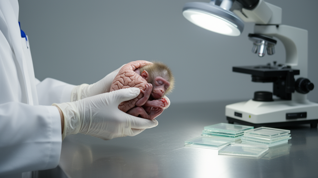
Every pregnancy unfolds inside an environment. The air a mother breathes, the metals in her blood, the viruses she encounters, the heat she endures, and the substances she consumes all reach the fetus through the placenta, and few tissues are as sensitive to these arrivals as the rapidly developing brain. A new review published in Advances in Drug and Alcohol Research argues that one of the best tools for understanding this vulnerability has been quietly underused: the baboon, Papio hamadryas, a nonhuman primate whose pregnancy, placenta, and fetal brain development mirror our own with a fidelity that rodents cannot match.
The review, led by researchers who have spent years working with pregnant baboons, surveys the milestones of prenatal brain maturation in nonhuman primates and catalogs how environmental insults—air pollution, ionizing radiation, toxic metals, pesticides, viral and bacterial infections, excess heat, and drugs of abuse—can derail them. The stakes are considerable. The fetal brain undergoes its most dramatic expansion during gestation, laying down the cortical layers, cerebellar circuits, and vascular networks that will support cognition and behavior for a lifetime. Disruptions during this window can produce long-term cognitive, emotional, and behavioral deficits, and identifying the moments of greatest sensitivity is central to prevention.
What makes the baboon so valuable is a matter of timing and anatomy. Rodents are altricial animals: their brains at birth correspond roughly to a human fetus in the third trimester, so many developmental processes that occur before birth in people happen after birth in mice and rats. In baboons and humans, by contrast, key steps of cerebellar development unfold during gestation itself. The external granule cell layer of the cerebellum, a proliferative zone that produces the granule neurons governing motor control and motor learning, can be tracked in fetal baboon tissue using molecular markers such as Pax6, which labels progenitor cells; Ki67, which marks dividing cells; and Tag1, which flags neurons that have begun to differentiate. Quantifying these cell populations offers a direct readout of whether an environmental insult has disturbed cerebellar development—a process that in rodents would be largely postnatal and therefore harder to align with human pregnancy.
The parallels extend across the entire brain. Baboons share roughly 92 percent of their genomic sequence with humans, and like us they possess a gyrencephalic cortex, folded into gyri and sulci rather than smooth. Imaging studies show that baboon brain volume and cortical surface area are highly heritable, that the corpus callosum follows a developmental trajectory closely mirroring the human one, and that grey-to-white matter ratios, myelination, and cortical laminar development share strong temporal and structural similarities with our own species. Even the cerebellum’s molecular layer thickens in late gestation and early infancy in both baboons and humans, reflecting comparable maturation of Purkinje cells.
The vascular system tells a similar story. By gestational day 120, corresponding to the end of the human second trimester, the four major cerebral arteries—anterior, middle, posterior, and basilar—are already distinguishable in the baboon fetus. By day 165, the third-trimester equivalent, fetal cerebral arteries can constrict in response to elevated intraluminal pressure, demonstrating myogenic tone, and they respond robustly to depolarizing stimuli such as potassium chloride, showing that smooth muscle contractility is being established in utero. The neurovascular unit—neurons, astrocytes, and endothelial cells—is remarkably conserved across primates, and primate brains arrive at birth with more advanced vascular networks than rodents, supporting the prolonged cortical development, including postnatal neurogenesis and gyrification, that characterizes our species.
Against this backdrop, the review assembles the evidence on environmental threats. Heavy metals cross the placental barrier with ease: arsenic is associated with miscarriage, stillbirth, and intrauterine growth restriction through oxidative stress and endocrine disruption; cadmium with preterm birth and small-for-gestational-age infants, possibly by interfering with placental nutrient transport; lead with low birth weight and impaired neurodevelopment; and methylmercury, accumulated from contaminated seafood, with lasting cognitive and motor deficits. Notably, a study of pregnant baboons measured forty elements across maternal and fetal blood, hair, nails, placenta, amniotic fluid, and fetal tissues, and found that element concentrations closely mirrored those of late-stage human pregnancies—reinforcing the baboon’s value for studying both normal maternal-fetal physiology and toxicology.
Physical and biological insults leave their own signatures. Excessive doses of ionizing radiation, above 0.1 gray, can cause miscarriage, growth restriction, malformations, and reduced cognitive function, with sensitivity depending on gestational stage; baboon studies have identified biomarkers such as aspartate aminotransferase, LDH, Flt3-ligand, and the neutrophil-to-lymphocyte ratio that distinguish total-body from partial-body irradiation and could guide treatment after accidental exposure. Maternal hyperthermia in pregnant baboons—body temperature of 41 to 42 degrees Celsius without fever—produced fetal blood pressure drops, elevated heart rate, severe acidosis, hypoxia, and hypercapnia, and doubled uterine contractility, raising the risk of preterm birth. Viral infections tell perhaps the starkest story: in baboons, herpesvirus papio 2 and cytomegalovirus cause placentitis with seroprevalence reaching 95 percent, and perinatal Zika virus infection produces significant fetal cerebral cortical injury that can end in fetal death.
Alcohol receives particularly detailed attention, and the baboon data are sobering. Three episodes of maternal alcohol exposure during the second-trimester equivalent, producing maternal blood alcohol levels around 80 milligrams per deciliter—the legal driving limit in most of the United States—caused fetal growth restriction, with amniotic fluid levels reaching 63 milligrams per deciliter, near the threshold of noticeable behavioral impairment in humans. Near term, exposed offspring showed reduced abdominal and head circumferences. A single binge episode, modeled with a gastro-nasal catheter delivering 3 grams of ethanol per kilogram to approximate a blood alcohol concentration of 0.2 percent—the equivalent of six to eight drinks in two hours—increased placental permeability and fetal brain perfusion, paradoxically making the fetal brain more vulnerable to toxic insult. Moderate doses during the second trimester dilated the middle cerebral artery, an effect reversed by blocking cannabinoid CB1 and CB2 receptors, and proteomic profiling revealed lasting alterations in mitochondrial and cytoskeletal proteins of cerebral arteries exposed earlier in gestation.
Cannabinoids and tobacco complete the picture. Roughly 5 percent of pregnant women self-medicate with marijuana, and chronic prenatal THC exposure in rhesus macaques altered fetal brain growth on MRI, with cerebrospinal fluid microRNAs pointing to disrupted axonal guidance and netrin signaling. Quantitative PCR in fetal baboon cerebellum confirms that both cannabinoid receptor genes, Cnr1 and Cnr2, are expressed at gestational day 120, the end of the second-trimester equivalent—meaning the fetal endocannabinoid system, which regulates synaptogenesis and circuit maturation, is present and potentially disruptable exactly when THC arrives. Epigenetic changes after prenatal cannabinoid exposure may even persist across generations, and are linked to psychiatric conditions including autism spectrum disorder, ADHD, and schizophrenia. Perinatal exposure to environmental tobacco smoke in rhesus monkeys selectively upregulated nicotinic acetylcholine receptors in the brainstem and cortex, a hallmark of nicotine neuroteratogenesis, while prenatal alcohol exposure in macaques and vervet monkeys reduced hippocampal and cortical neuron numbers and disrupted miRNA regulation of genes critical for synaptic development, including a previously unknown link to the EFNB1 gene.
The authors acknowledge that nonhuman primate research carries significant ethical and logistical burdens—long lifespans, complex care, strict welfare requirements—but they argue the translational payoff justifies the investment. Rodent preclinical results frequently fail in human clinical trials, a gap that may stem from precisely the developmental and vascular differences the baboon model is positioned to resolve. Computational models already predict that primates, including humans, are more sensitive to ethanol’s inhibition of neocortical neuronal proliferation than rodents, with significant neuronal deficits at blood alcohol concentrations equivalent to a single standard drink within an hour. As cannabis legalization reshapes perceptions of safety, as air pollution and heat extremes intensify, and as viral threats continue to emerge, the baboon offers a rare window into the mechanisms by which the prenatal environment writes itself into the developing brain—and a foundation for the prevention and treatment strategies that neurodevelopmental medicine urgently needs.
Subject of Research: Prenatal environmental exposures and fetal brain development studied with baboon and other nonhuman primate models
Article Title: Prenatal environmental exposures and brain development: studies with baboons and other nonhuman primates
Article References: Iskusnykh, I. Y., Thapa, S., Chizhikov, V. V., & Bukiya, A. N. (2025). Prenatal environmental exposures and brain development: studies with baboons and other nonhuman primates. Advances in Drug and Alcohol Research, 5, Article 14858. https://doi.org/10.3389/adar.2025.14858
Image Credits: AI Generated
Keywords: neuroscience, developmental biology, toxicology, baboon model, fetal brain, prenatal exposure, FASD, cannabinoids, Prenatal, environmental, exposures, brain
Cite Scienmag News
Cassandra Pierce. (October 16, 2026). Baboon pregnancies reveal how pollution, infection and alcohol reshape the fetal brain. Scienmag. https://scienmag.com/baboon-pregnancies-reveal-how-pollution-infection-and-alcohol-reshape-the-fetal-brain/
Cassandra Pierce. "Baboon pregnancies reveal how pollution, infection and alcohol reshape the fetal brain." Scienmag, 16 October 2026, https://scienmag.com/baboon-pregnancies-reveal-how-pollution-infection-and-alcohol-reshape-the-fetal-brain/. Accessed 16 October 2026.
Cassandra Pierce. "Baboon pregnancies reveal how pollution, infection and alcohol reshape the fetal brain." Scienmag. October 16, 2026. https://scienmag.com/baboon-pregnancies-reveal-how-pollution-infection-and-alcohol-reshape-the-fetal-brain/


