


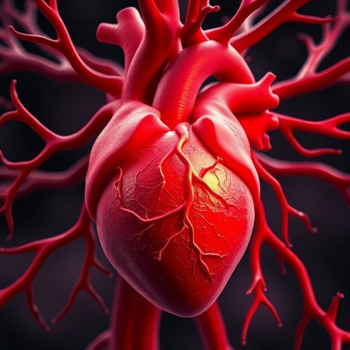
Coronary artery disease has long been understood as a problem of obstruction: when a major vessel narrows or becomes blocked, blood can no longer reach the heart muscle efficiently, increasing the risk of ischemia, heart attack and permanent tissue damage. Yet the heart is not entirely dependent on its primary coronary arteries. It can develop small alternative routes known as collateral arteries, which redirect blood around an obstruction and help preserve the function of vulnerable cardiac tissue. New research in genetically engineered mice now challenges a central assumption about how these natural bypass vessels form. Rather than arising mainly through the enlargement of pre-existing arterial connections, many new coronary collaterals appear to be built primarily from capillaries, the smallest vessels in the circulatory system.
The study, led by Mingjun Zhang and colleagues and published in Science, provides evidence that capillary endothelial cells can undergo a substantial change in identity and behavior during cardiac repair. Endothelial cells form the inner lining of blood vessels, but they are not a uniform population. Cells lining arteries, veins and capillaries carry distinct molecular signatures and respond differently to changes in blood flow, oxygen availability and growth signals. By following these populations during the development of coronary collaterals, the researchers found that capillary endothelial cells contributed extensively to newly formed arterial vessels. The result suggests that the heart can generate arterial bypass routes through a developmental process that is more flexible than previously recognized.
The traditional explanation for collateral formation has focused on arteriogenesis, a process in which small arterial connections that already exist between neighboring vascular territories enlarge after a blockage. When blood is forced through these narrow channels, the resulting changes in fluid shear stress stimulate endothelial cells and surrounding vascular smooth muscle cells. In response, the vessels remodel, expand and acquire characteristics of larger arteries. This mechanism is important in many settings, but it does not fully explain the appearance of collateral vessels in regions where mature arterial connections are absent or insufficient. Earlier studies had begun to suggest that new vessels could also arise de novo, but the precise cellular sources of these arteries remained uncertain.
To resolve that question, Zhang and the team used complementary genetic lineage-tracing strategies in mice. These tools allow researchers to label a defined endothelial-cell population and then determine where its descendants appear as tissues develop or respond to injury. The investigators tracked cells associated with different parts of the coronary vascular network, including arterial and capillary endothelium. By comparing the distribution of lineage-marked cells before and after coronary obstruction, they were able to determine which populations were incorporated into newly developing collateral arteries. The dual tracing approach was particularly important because it reduced the risk of attributing vessel formation to a single cell type based solely on molecular markers, which can change during vascular remodeling.
The results indicated that capillary endothelial cells were major contributors to de novo coronary collateral formation. Following the loss or restriction of blood flow through a coronary artery, capillary-derived cells began expressing features associated with arterial endothelium and became incorporated into larger, conduit-like vessels. This was not simply a passive enlargement of existing capillaries. It involved a coordinated conversion in which cells changed their gene-expression programs, reorganized their structure and participated in the construction of vessels capable of carrying blood across an area affected by the blockage. In effect, the capillary network served as a reservoir of cells that could be recruited to build a new arterial route when the heart required an alternative supply.
The researchers also identified a role for signaling driven by vascular endothelial growth factor, or VEGF, in promoting the capillary-to-artery transition. VEGF is widely known for stimulating blood-vessel growth, particularly under conditions of low oxygen or tissue stress. Its effects, however, depend on the cellular context, the receptors involved and the broader signaling environment. In this study, VEGF-related signaling helped activate the molecular changes required for capillary endothelial cells to acquire arterial characteristics. The findings suggest that VEGF does more than encourage the sprouting of new microvessels; under appropriate conditions, it may also help determine the identity and functional organization of the vessels that emerge.
This distinction could be important for future therapeutic strategies. Simply increasing the number of small blood vessels may not be enough to restore circulation to damaged heart muscle. A useful repair response must produce vessels that connect effectively with the existing coronary network, withstand arterial pressure and deliver a sustained flow of oxygenated blood. The capillary-derived collaterals described in the study appear to represent a process directed toward that larger-scale vascular architecture. If the relevant VEGF-dependent pathway can be controlled precisely, it might eventually be possible to encourage the formation of functional collateral arteries after a coronary event, potentially reducing the amount of tissue lost to ischemia.
The findings remain experimental and do not yet establish that the same process can be safely or effectively induced in people. Mouse hearts differ from human hearts in anatomy, physiology and the organization of their coronary circulation. VEGF-based treatments also present challenges because excessive or poorly localized signaling can produce abnormal vessels, edema or unwanted vascular growth. Any future therapy would therefore need to regulate the timing, intensity and location of the response rather than simply deliver a large dose of a growth factor. Researchers will also need to determine how the newly formed vessels mature, connect to the wider circulation and perform over the long term after injury.
For patients with coronary artery disease, the immediate standards of care remain prevention, medication, catheter-based intervention and bypass surgery when clinically indicated. These approaches can restore blood flow, but they are invasive or limited by the complexity of the disease and the condition of the patient. A treatment that could activate the heart’s own capacity to create collateral arteries would offer a different form of repair, particularly for people whose vessels are too diffuse or fragile for conventional revascularization. The new study provides a cellular explanation for how such a response may begin: capillary endothelial cells can be redirected toward an arterial fate, and VEGF-linked signaling appears to help guide that transformation. By revealing the source of the cells and the pathway involved, the work moves the field closer to designing therapies that support the heart’s natural vascular repair system.
News Publication Date: 20-Aug-2026
Web References: https://doi.org/10.1126/science.ady3027
References: Zhang et al., “Tracing the origins of de novo coronary collateral formation in cardiac repair,” Science, DOI: 10.1126/science.ady3027.
Cite Scienmag News
Drew Townsend. (August 21, 2026). Heart backup blood vessels grow from capillaries—not arteries. Scienmag. https://scienmag.com/heart-backup-blood-vessels-grow-from-capillaries-not-arteries/
Drew Townsend. "Heart backup blood vessels grow from capillaries—not arteries." Scienmag, 21 August 2026, https://scienmag.com/heart-backup-blood-vessels-grow-from-capillaries-not-arteries/. Accessed 3 September 2026.
Drew Townsend. "Heart backup blood vessels grow from capillaries—not arteries." Scienmag. August 21, 2026. https://scienmag.com/heart-backup-blood-vessels-grow-from-capillaries-not-arteries/


