


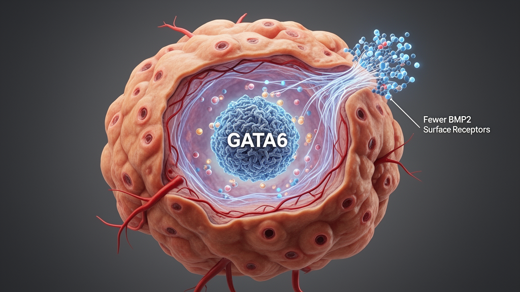
Elevated bone morphogenetic protein 2 (BMP2) in lung squamous cell carcinoma (LUSC) was associated with poorer survival in a study combining tissue analysis, genetic experiments in cell lines, mouse xenografts and molecular assays. The researchers found that the transcription factor GATA6 directly suppresses BMP2 expression, while BMP2 promotes proliferation, migration and invasion of tumor cells by activating the MAPK–ERK1/2 signaling pathway. The results identify a regulatory relationship between GATA6 and BMP2, but the findings remain preclinical and the prognosis data were not fully consistent across public cohorts.
Non-small cell lung cancer accounts for approximately 85% of lung cancer cases, and about 30% of these cases are LUSC. Although targeted therapy and immunotherapy have produced major advances in non-small cell lung cancer, LUSC remains difficult to treat with current traditional approaches. BMP2 has important functions in embryonic development and lung formation, but its role in cancer is context dependent. The source study notes that BMP2 can suppress cancer stem-cell expansion and induce osteogenic differentiation in some settings, while also functioning as an oncogene by regulating cancer-related genes. Earlier reports found elevated BMP2 in non-small cell lung cancer and an association between high serum BMP2 and poor survival, but its effects and regulatory mechanisms in LUSC required further investigation.
The investigators first combined five Gene Expression Omnibus datasets to search for genes associated with LUSC tumorigenesis or metastasis. Comparisons included tumor tissue against normal bronchial biopsies or tumor-free lung, as well as the KLN205 LUSC cell line and its metastatic subclone, KLN.LN4K1. Intersection of the differentially expressed genes identified BMP2, GAPDH and LIFR as common candidates. Analysis of The Cancer Genome Atlas through GEPIA2 associated high BMP2 expression with unfavorable survival. The researchers then examined protein expression in a tissue microarray, finding significantly higher BMP2 immunohistochemistry scores in LUSC tissue than in normal lung and in paired adjacent normal tissues.
Patients classified as having high BMP2 expression, defined by an immunohistochemistry score above the cohort mean, had shorter survival than those with low expression. High BMP2 was also associated with positive lymph-node status and higher N stage. In univariate and multivariable analyses that included tumor size, lymph-node status, histological grade and BMP2 expression, high BMP2 remained a risk factor for prognosis; the reported multivariable hazard ratio was 3.032, with a 95% confidence interval of 1.098 to 8.372. The tissue-microarray survival comparison included 42 patients per group. The association was not consistent in every public dataset: an analysis of 780 patients with LUSC in Kaplan–Meier Plotter found no significant relationship between BMP2 expression and survival, with a hazard ratio of 1.09 and p value of 0.39. Differences in cohort composition and platforms may have contributed to the discrepancy, and serum BMP2 was not measured in the study.
To test the effect of BMP2 directly, the team generated stable BMP2-overexpressing LUSC cell lines. In NCI-H2170 cells, BMP2 overexpression increased growth rates, the proportion of cells incorporating EdU during DNA synthesis, and migration and invasion in transwell assays. The same pattern appeared in NCI-H226 cells: elevated BMP2 was associated with faster growth, more EdU-positive cells and greater migratory and invasive capacity. Loss-of-function experiments produced the converse result. After the researchers depleted BMP2 in SK-MES-1 cells, those cells grew more slowly and showed reduced migration and invasion. Together, the complementary gain- and loss-of-function experiments support a functional role for endogenous as well as experimentally increased BMP2 in malignant cell behavior.
The effects were also examined in vivo. Subcutaneous implantation of BMP2-overexpressing NCI-H2170 cells into athymic nude mice produced xenograft tumors with significantly greater volume and weight than tumors formed by control cells, with eight animals per group. A separate tail-vein injection model provided preliminary evidence about metastasis. Bioluminescent signals appeared in the lung, liver and brain of three of ten mice receiving BMP2-overexpressing cells, compared with one of ten receiving controls; the control signal was confined to the lung. A brain lesion from a BMP2-expressing mouse was confirmed by hematoxylin and eosin staining. The difference in metastatic incidence did not reach statistical significance because of the small number of events, so the result suggests a possible increase in metastatic potential rather than establishing it.
The researchers next searched for transcription factors capable of controlling BMP2. Intersecting predictions from KnockTF, AnimalTFDB, CistromeDB and GTRD produced 10 candidates, including MYC and RELA. A separate comparison of GATA6-associated expression changes identified BMP2 as a potential target. In LUSC cells, forcing GATA6 expression reduced BMP2 messenger RNA and protein, whereas GATA6 depletion increased BMP2 messenger RNA. Promoter analyses predicted seven GATA6-binding regions within the 3,000-base-pair region upstream of the BMP2 gene. Reporter assays showed that GATA6 inhibited BMP2 promoter activity, and mutation of individual predicted sites identified regions at −793 to −784, −1286 to −1273 and −1717 to −1708 base pairs as functionally important.
Chromatin immunoprecipitation followed by polymerase chain reaction in NCI-H2170 cells showed enrichment of the BMP2 promoter around all three functional sites in anti-GATA6 immunoprecipitates relative to immunoglobulin G controls, with no enrichment at a negative control region. These results provide direct evidence that GATA6 occupies the BMP2 promoter in the native chromatin of the cancer cells. The mechanism is not universal: the source article cites earlier work in which GATA6 increased, rather than reduced, BMP2 promoter activity in an adrenocortical cancer line. The researchers suggest that differences in co-regulator recruitment or promoter epigenetics may account for this tissue-dependent behavior, a question that would require chromatin profiling and comparisons of GATA6-associated proteins across cell types.
Functional experiments connected this transcriptional control to tumor behavior. The authors had previously identified GATA6 as a tumor suppressor in LUSC. When GATA6 was overexpressed by itself, LUSC cells grew less rapidly and showed reduced migration and invasion, but simultaneous BMP2 overexpression counteracted those suppressive effects. RNA sequencing of BMP2-overexpressing NCI-H2170 cells identified 156 upregulated and 111 downregulated genes at a fold-change threshold greater than two. Pathway analysis pointed to the non-canonical MAPK route, and immunoblotting showed increased phosphorylation of ERK1/2 without a significant rise in phosphorylated SMAD1/5/8/9. BMP2 also increased expression of MMP9, a protein implicated in extracellular-matrix remodeling, while genes associated with extracellular-matrix organization were prominent among the affected pathways.
Treatment of BMP2-overexpressing NCI-H2170 cells with the MEK inhibitor U0126 reduced the elevated ERK1/2 phosphorylation and abolished the increased proliferation and migration, bringing both phenotypes to levels comparable with controls. The inhibitor experiments support the necessity of MEK/ERK signaling for the BMP2-driven phenotypes in this cellular model, rather than merely showing an association between pathway activity and growth. Overall, the study proposes a sequence in which reduced GATA6 transcriptional repression permits BMP2 elevation, BMP2 activates non-canonical ERK1/2 signaling and alters extracellular-matrix-related programs, and these changes support proliferation and invasion. The evidence comes mainly from established cell lines and immunodeficient mouse models; orthotopic tumors and genetically engineered models would better reproduce the human lung environment. The work therefore identifies a mechanistic target for further study, not an established treatment or a clinically validated prognostic biomarker for LUSC.
Subject of Research: Molecular regulation of lung squamous cell carcinoma by the GATA6–BMP2–ERK1/2 pathway
Article Title: BMP2, transcriptionally regulated by GATA6, accelerates the proliferation and invasion of lung squamous carcinoma cells
Article References: Yang, H., Huang, C., Wang, H., & Chen, L. (2026). BMP2, transcriptionally regulated by GATA6, accelerates the proliferation and invasion of lung squamous carcinoma cells. iScience, 29(11), Article 117882. https://doi.org/10.1016/j.isci.2026.117882
Image Credits: AI Generated
DOI: 10.1016/j.isci.2026.117882
Keywords: lung squamous cell carcinoma, BMP2, GATA6, ERK1/2 signaling, MAPK pathway, cancer-cell proliferation, tumor invasion, mouse xenografts, transcriptional regulation, transcriptionally, regulated, accelerates
Cite Scienmag News
Nathaniel Bowman. (October 20, 2026). GATA6 represses BMP2 to restrain lung squamous cell carcinoma. Scienmag. https://scienmag.com/gata6-represses-bmp2-to-restrain-lung-squamous-cell-carcinoma/
Nathaniel Bowman. "GATA6 represses BMP2 to restrain lung squamous cell carcinoma." Scienmag, 20 October 2026, https://scienmag.com/gata6-represses-bmp2-to-restrain-lung-squamous-cell-carcinoma/. Accessed 20 October 2026.
Nathaniel Bowman. "GATA6 represses BMP2 to restrain lung squamous cell carcinoma." Scienmag. October 20, 2026. https://scienmag.com/gata6-represses-bmp2-to-restrain-lung-squamous-cell-carcinoma/


