


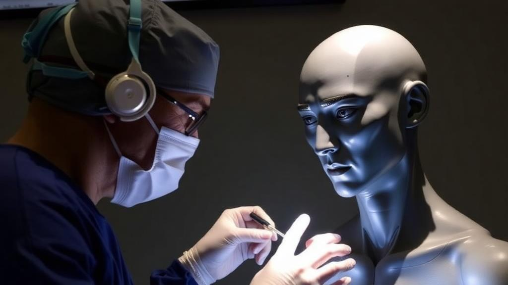
Under an operating microscope, the difference between success and failure can be measured in fractions of a millimeter. A surgeon stitching together a vessel thinner than a strand of spaghetti must place each suture with a precision that leaves no room for improvisation. This is the world of microsurgery, the cornerstone of modern reconstructive surgery, and a new narrative review published in BMC Plastic and Reconstructive Surgery traces how surgeons have learned to master it, from the apprenticeship benches of the 1960s to today’s virtual reality simulators and smartphone-based training kits. The review, authored by Hatan Mortada of King Saud University in Riyadh and published on 3 October 2025, synthesizes six decades of educational innovation and finds a field in the midst of a profound transformation.
The story begins with two pioneers. In 1960, Julius Jacobson reported the first microvascular anastomosis performed under an operating microscope, a technical breakthrough that made it possible to join blood vessels far too small for the naked eye. Four years later, Harry Buncke demonstrated the successful replantation of a rabbit ear using microminiature vascular anastomoses, a feat that electrified the surgical world and showed what the technique could ultimately achieve in patients. These milestones laid the groundwork for modern microsurgery, but they also established a training culture that would persist for decades: learning by doing, under the eye of a mentor, in the operating room itself.
That apprenticeship model had undeniable strengths. Trainees gained proficiency through repeated exposure and direct feedback, practicing vascular anastomoses on live animal models such as rats and rabbits with basic equipment like magnifying loupes and early operating microscopes. By the 1970s, microsurgery had gained traction in plastic surgery and neurosurgery, and institutions such as the University of California, San Francisco, under Buncke’s leadership, became key centers of education. Yet access remained strikingly limited. By the early 1980s, fewer than ten dedicated microsurgical training centers existed worldwide, most of them concentrated in North America and Europe. High equipment costs, scarce mentors, and limited access to models meant that the steep learning curve of microsurgery was climbed by only a privileged few.
The forces that shattered this old order were administrative as much as scientific. In 2003, the Accreditation Council for Graduate Medical Education formalized reduced resident duty hours in the United States, sharply curtailing the time trainees could spend in the operating room. At the same time, heightened patient safety standards made it less acceptable for trainees to learn fundamental skills on live patients. Educators were compelled to find alternatives, and the result was a wave of structured courses, simulation laboratories, and formal curricula designed to build competency before a trainee ever touched a patient. Programs such as the Columbia University Microsurgical Training Course and the Mayo Clinic Microsurgery Course combined didactic lectures with hands-on practice to establish baseline skills outside the clinical environment.
The evidence that such training works has accumulated steadily. A systematic review by Ghanem and colleagues found that low-fidelity laboratory models effectively improve microsurgical technical skills, and that these skills transfer to higher-fidelity models and live tissue performance. Perhaps more striking is the Ludwigshafen concept from Germany, where Boecker and colleagues showed that a structured microsurgical curriculum supported by a 24-hour accessible training facility significantly reduced complication rates and operative times in lower extremity free flap surgeries performed by residents. Mentorship still matters within these systems: Paladino and colleagues compared microsurgery courses with and without dedicated expert instruction and found that students guided by experts achieved superior improvements in patency rates, anastomotic quality, and procedural timing.
At the high-fidelity end of the spectrum sit wet labs, long regarded as the gold standard for realistic practice. Trainees anastomose rat femoral arteries, gaining live-tissue handling experience with genuine physiological feedback under the supervision of experienced microsurgeons. But live animal models raise ethical and logistical concerns, and recent research has shown these can be addressed without sacrificing quality. In a randomized noninferiority trial, Esanu and colleagues demonstrated that spacing live rat sessions every eight weeks instead of every four or six, while supplementing with lower-fidelity models, did not compromise skill acquisition. Lahiri and colleagues similarly found that cutting the number of live rats per participant from five to as few as three, while increasing time on synthetic and ex vivo models, achieved comparable patency rates and procedural times. Institutions have even developed portable wet lab setups so visiting trainees outside major academic centers can access these experiences.
The most viral chapter of this evolution, however, belongs to the digital revolution. Virtual reality platforms offer immersive, risk-free environments where trainees can repeat complex tasks such as microvascular anastomosis endlessly, receiving real-time feedback and performance metrics without any biological material. While microsurgery-specific trials remain limited, the precedent from laparoscopic surgery is dramatic: in a randomized controlled trial, VR-trained residents performed procedures 29 percent faster and made six times fewer errors than their traditionally trained counterparts. Alongside VR, a flourishing ecosystem of low-cost tools has emerged. Couceiro and colleagues described a practical model using chicken thighs and wings that simulates clinical conditions better than inert synthetics. Navia and colleagues validated MicrosimUC, a do-it-yourself kit with a modified miniature microscope that produced skill gains comparable to formal laboratory courses. De Fré and colleagues even showed that a smartphone-based training model, despite lacking stereoscopic depth, effectively improved anastomosis quality, patency, and procedural timing. Video-based learning, too, has proven non-inferior to traditional tutor-led courses for novices.
Objective measurement is the newest frontier. Lyon and colleagues applied three-dimensional motion-tracking technology to analyze hand and instrument movements during microsurgical anastomoses, showing that metrics such as path length, idle time, and working volume can objectively differentiate trainees from experts, a promising basis for competency-based feedback. Chauhan and colleagues developed the Fundamentals of Microsurgery curriculum, adapting validated laparoscopic education principles with stepwise tasks from basic instrument handling to complex vessel anastomosis in confined spaces; progression through it translated into improved intraoperative performance, reduced mental workload, and decreased performance anxiety. Joy and colleagues introduced a high-fidelity platform using cryopreserved human veins and a pulsatile flow circuit, boosting resident confidence over repeated sessions. Yet a note of caution runs through the literature: in a systematic review by Javid and colleagues, only a handful of the 64 identified microsurgical simulation models achieved high levels of recommendation, with most lacking rigorous psychometric validation, a gap that undermines the generalizability of VR and other tools and slows their integration into formal curricula and credentialing.
The global picture remains uneven. Surveys of U.S. integrated plastic surgery programs found that while 94 percent had access to a training microscope, 78 percent lacked a formal curriculum. A 2023 review of 103 U.S. programs found only 35 percent offered a formal microsurgery curriculum, and fewer than one-fourth included benchmark assessments. Internationally, centers in South Korea, India, and Taiwan embed simulation directly into active surgical rotations, while organizations such as the American Society for Reconstructive Microsurgery, the World Society for Reconstructive Microsurgery, and the digitally native International Microsurgery Club, with more than 20,000 members, work to standardize training across borders. Disparities in resource access, particularly in low-income regions, still limit adoption of VR and wet labs. Looking ahead, the review points to artificial intelligence-powered adaptive simulators and telesimulation connecting remote learners with experts as the next frontier. The through-line of six decades is clear: the mentor’s steady hand remains irreplaceable, but it is now amplified by silicon, simulation, and a global network, ensuring that the artistry of microsurgery can be taught to anyone, anywhere.
Subject of Research: The evolution of microsurgical training methods from apprenticeship to simulation-based education
Article Title: The evolution of microsurgery training: a narrative review of historical foundations and modern innovations
Article References: Mortada, H. (2025). The evolution of microsurgery training: a narrative review of historical foundations and modern innovations. BMC Plastic and Reconstructive Surgery, 1(1), Article 3. https://doi.org/10.1186/s44452-025-00002-8
Image Credits: AI Generated
DOI: 10.1186/s44452-025-00002-8
Keywords: microsurgery, surgical training, virtual reality, simulation, medical education, wet labs, mentorship, reconstructive surgery, residency, surgical curriculum, telemedicine, history of medicine
Cite Scienmag News
Ophelia Keating. (October 2, 2026). From Apprentice to Avatar: How Microsurgery Training Entered the Digital Age. Scienmag. https://scienmag.com/from-apprentice-to-avatar-how-microsurgery-training-entered-the-digital-age/
Ophelia Keating. "From Apprentice to Avatar: How Microsurgery Training Entered the Digital Age." Scienmag, 2 October 2026, https://scienmag.com/from-apprentice-to-avatar-how-microsurgery-training-entered-the-digital-age/. Accessed 4 October 2026.
Ophelia Keating. "From Apprentice to Avatar: How Microsurgery Training Entered the Digital Age." Scienmag. October 2, 2026. https://scienmag.com/from-apprentice-to-avatar-how-microsurgery-training-entered-the-digital-age/


