


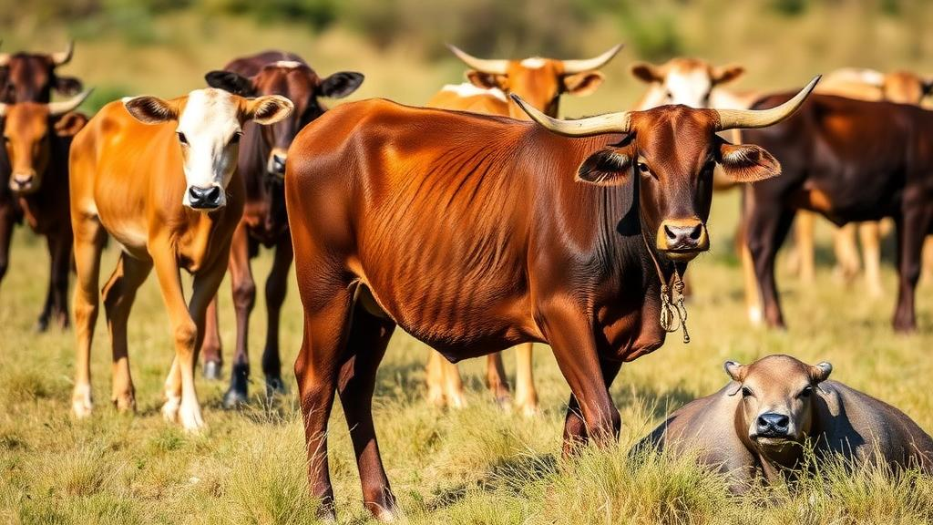
On the grasslands of Ol Pejeta Conservancy in central Kenya, cattle and wild grazers share pasture, water and, as a new study shows, a far richer community of parasitic worms than conventional diagnostics have ever revealed. Researchers led by Erhan Yalcindag of the University of Edinburgh’s Roslin Institute, working with colleagues at the International Livestock Research Institute and the conservancy itself, applied a deep-amplicon sequencing technique known as nemabiome metabarcoding to cattle at this wildlife–livestock interface. Their results, published in Parasites & Vectors, document eight gastrointestinal nematode species circulating in a single herd and demonstrate that mixed infections, far from being the exception, are the overwhelming norm. The work offers one of the most detailed molecular pictures yet of worm biodiversity in African livestock, and it arrives at a moment when the tools used to fight these parasites are struggling to keep pace with their evolution.
Gastrointestinal nematodes are among the most economically consequential pathogens of grazing livestock worldwide. In cattle, species such as Haemonchus placei, a blood-feeding abomasal worm, and various Cooperia species that damage the small intestine cause weight loss, reduced milk yield, poor fertility and, in heavy infections, death. The global cost is measured in billions of dollars annually, and the burden falls disproportionately on smallholder and pastoralist communities across sub-Saharan Africa, where livestock are both income and insurance. Yet the epidemiology of these worms in Africa remains strikingly under-characterised. Most surveillance still depends on faecal egg counts, which estimate the number of eggs shed per gram of faeces, and on the microscopic identification of third-stage larvae cultured from faecal samples. Both approaches have fundamental limitations that the new study set out to address.
Faecal egg counts cannot distinguish between species, because the eggs of many strongylid nematodes are morphologically near-identical. Larval culture and identification improve on this, but the technique is laborious, requires skilled parasitologists, takes one to two weeks, and is subject to culture biases that can distort the apparent composition of an infection. Different species develop and survive in culture at different rates, meaning the larval population recovered in a petri dish may not faithfully represent the worm population inside the animal. In tropical settings, where ambient temperatures accelerate larval development and where laboratory infrastructure may be limited, these problems are compounded. The result is a sparse and potentially skewed picture of nematode diversity across much of the continent, precisely where that diversity matters most for designing effective control.
The nemabiome approach sidesteps these constraints by reading the parasites’ DNA directly. The method targets the internal transcribed spacer-2 region, or ITS-2, a stretch of ribosomal DNA that varies sufficiently between species to serve as a barcode. Using primers that amplify this region from any Clade V nematode, the strongylid group that includes the major livestock worms, researchers can sequence thousands of amplicons in parallel from a single sample. Bioinformatic pipelines then sort the resulting reads into species-level operational taxonomic units and estimate their relative abundances. Because the technique works on any life stage containing usable DNA, it can in principle be applied to eggs in faeces, to first-stage larvae hatched from those eggs, or to third-stage larvae recovered from culture, allowing researchers to compare what each sample type reveals.
That is exactly what the team did at Ol Pejeta, a conservancy where livestock grazing is deliberately integrated with wildlife conservation, including populations of elephants, rhinos and large predators. The study focused on adult cows and their calves, sampling faeces, first-stage larvae and cultured third-stage larvae from individual animals. The nemabiome analysis confirmed infections with eight species: Cooperia curticei, Cooperia pectinata, Cooperia punctata, Haemonchus placei, Haemonchus contortus, Teladorsagia circumcincta, Trichostrongylus axei and Trichostrongylus colubriformis. Several of these findings are notable in themselves. Haemonchus contortus, classically a parasite of sheep and goats, was detected in cattle, raising questions about cross-species transmission at the interface. Teladorsagia circumcincta, another small-ruminant worm, likewise appeared in the cattle nemabiome, a signal that would have been easy to miss with morphology-based methods alone.
Perhaps the most striking quantitative finding concerns coinfection. Mixed-species infections accounted for 77.4 percent of all infections detected in the study animals. In other words, the typical infected cow at Ol Pejeta was not carrying a single worm species but a community of them, with the composition varying from animal to animal. This matters because nematode species differ in their pathogenicity, their seasonal dynamics and, critically, their response to anthelmintic drugs. A treatment regimen informed only by faecal egg counts treats the infection as an undifferentiated mass; a regimen informed by metabarcoding can recognise, for example, that a Haemonchus-dominated infection may demand different management than one dominated by Trichostrongylus. The study’s authors argue that uncovering this diversity opens new opportunities to investigate interactions between worm species within the host, including competition, facilitation and their consequences for disease severity and drug efficacy.
Age emerged as a second axis of variation. Calves carried higher infection rates than their dams and harboured more diverse nematode communities. This pattern is consistent with the immunology of helminth infection: adult cattle gradually acquire partial immunity through repeated exposure, which suppresses worm establishment and fecundity, whereas young animals, still immunologically naive, accumulate infections more freely. From a control perspective, the finding reinforces the importance of monitoring calves specifically, since they are both the most heavily parasitized cohort and the one in which subclinical damage to gut and abomasum can permanently compromise growth. It also suggests that dam–calf pairs provide a natural experiment for studying the development of nemabiome diversity over time, a direction the authors see as promising for future work.
Equally consequential is the study’s practical demonstration of which sample types can carry the analysis. Faecal samples and first-stage larvae both proved reliable sources of nematode DNA, yielding species compositions consistent with the gold-standard cultured third-stage larvae. This is a significant result for tropical field settings. Third-stage larval culture requires incubation, careful moisture and temperature management, and weeks of waiting; faeces require a bag and a courier. If routine surveillance can be run on faecal or first-stage larval DNA, the barrier to implementing molecular monitoring in African veterinary services drops dramatically. The authors position this as offering a practical alternative to labour-intensive L3 culturing, and the implication for national control programmes is clear: species-level surveillance could become as routine as the egg counts it would complement.
The wildlife–livestock interface setting adds a further layer of significance. Ol Pejeta’s cattle graze land shared with wild ungulates, and several of the nematode species detected are known to infect multiple host species. Haemonchus contortus, for instance, circulates in wild ruminants as well as small livestock, and Trichostrongylus species have broad host ranges. Molecular barcoding of livestock parasites therefore offers a window into transmission dynamics that span the domestic–wild boundary, something morphology-based diagnostics have never been able to resolve at scale. The authors argue that in such complex ecosystems, nemabiome approaches provide strong potential for unbiased disease surveillance, capturing the full parasite community rather than only the species a technician expects to find. As land-use change brings livestock and wildlife into ever closer contact across East Africa, tools that can track parasite flow between them will become increasingly valuable for both conservation and animal health.
The study also serves as a proof of concept for building African nemabiome reference data. Metabarcoding is only as good as its reference database; reads can be assigned to species only when that species’ ITS-2 sequence has been catalogued. By generating and validating species assignments in a Kenyan herd, the team has contributed to the baseline needed for wider deployment across the continent. Funded in part by the Bill & Melinda Gates Foundation and UK aid through the Centre for Tropical Livestock Genetics and Health, the work reflects a growing investment in molecular epidemiology for African livestock systems. The immediate next steps, the authors suggest, include exploiting the revealed diversity to study inter-species interactions within hosts and to track how nematode communities shift with season, treatment and grazing management. For a class of parasites that has long been counted but rarely identified, the era of seeing the whole community has begun.
Subject of Research: Gastrointestinal nematode diversity and coinfection in Kenyan cattle at a wildlife–livestock interface, assessed by nemabiome metabarcoding
Article Title: Gastrointestinal nematode diversity in Kenyan cattle at wildlife–livestock interfaces: a deep-amplicon sequencing approach
Article References: Yalcindag, E., Karani, B. E., Vasoya, D., van Bunnik, B. A. D., Freeman, E. J., Ngulu, S., van Aardt, R., Toye, P., Sargison, N., Morrison, L. J., & Bronsvoort, B. M. D. C. (2026). Gastrointestinal nematode diversity in Kenyan cattle at wildlife–livestock interfaces: a deep-amplicon sequencing approach. Parasites & Vectors. https://doi.org/10.1186/s13071-026-07657-x
Image Credits: AI Generated
DOI: 10.1186/s13071-026-07657-x
Keywords: gastrointestinal nematodes, nemabiome, metabarcoding, Kenya, cattle, wildlife–livestock interface, Haemonchus, Cooperia, coinfection, parasite surveillance, ITS-2 sequencing, livestock health
Cite Scienmag News
William Thompson. (October 2, 2026). DNA Sequencing Reveals Hidden Worm Coinfections in Kenyan Cattle Grazing Beside Wildlife. Scienmag. https://scienmag.com/dna-sequencing-reveals-hidden-worm-coinfections-in-kenyan-cattle-grazing-beside-wildlife/
William Thompson. "DNA Sequencing Reveals Hidden Worm Coinfections in Kenyan Cattle Grazing Beside Wildlife." Scienmag, 2 October 2026, https://scienmag.com/dna-sequencing-reveals-hidden-worm-coinfections-in-kenyan-cattle-grazing-beside-wildlife/. Accessed 4 October 2026.
William Thompson. "DNA Sequencing Reveals Hidden Worm Coinfections in Kenyan Cattle Grazing Beside Wildlife." Scienmag. October 2, 2026. https://scienmag.com/dna-sequencing-reveals-hidden-worm-coinfections-in-kenyan-cattle-grazing-beside-wildlife/


