


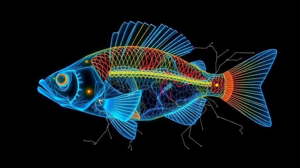
In a landmark study published in Nature, a team of neuroscientists led by Krista E. Perks, Mariela D. Petkova and Salomon Z. Muller, working across Columbia University, Harvard University and Google Research, has reconstructed the complete wiring diagram of a cerebellum-like brain structure in an electric fish, exposing in unprecedented detail how neural circuits learn to predict and cancel out the sensory consequences of an animal’s own behavior. The work, published under the title Connectome analysis of a cerebellum-like circuit for sensory prediction, combines nanoscale electron microscopy, cell-type-specific physiology and computational modeling to answer one of neuroscience’s most stubborn questions: how does a brain coordinate learning that is spread across many cell types and many layers of a network at once?
The animal at the center of the study is the mormyrid electric fish, a creature that generates its own weak electric field and senses the world through distortions of that field. Every time the fish fires its electric organ, it also sends a copy of the motor command, known as the corollary discharge, to a hindbrain structure called the electrosensory lateral line lobe, or ELL. This structure, architecturally reminiscent of the cerebellum, uses the corollary discharge to generate a prediction of the sensory input that the fish’s own discharge will produce. That prediction is then subtracted from the actual sensory stream, a process scientists call negative imaging. The result is elegant: predictable self-generated signals vanish, while unexpected signals, such as the electrical signature of nearby prey or a communicating neighbor, stand out in sharp relief.
What makes the fish such a powerful model is that this cancellation is learned continually. When the fish’s environment changes, for example when the amplitude of its electric organ discharge shifts, the circuit adjusts within minutes, and it does so without erasing previously learned predictions. This is a continual learning problem, the biological counterpart of a challenge that plagues artificial neural networks, where new training catastrophically overwrites old memories. Previous work from the same laboratories had shown that learning in the ELL is distributed across multiple synaptic sites, but the detailed wiring needed to explain how those sites cooperate had never been mapped.
To build that map, the team reconstructed a volume of the ELL at a resolution of 4 by 4 by 30 nanometers using serial section electron microscopy, a dataset generated in Jeff Lichtman’s laboratory at Harvard and segmented with automated tools developed at Google. The researchers painstakingly proofread and classified hundreds of neurons, including more than one hundred MG cells and nearly six hundred SG cells, tracing their axons and dendrites and annotating thousands of synapses. Every reconstructed connection was catalogued, allowing the team to ask not just which cells talk to which, but whether the pattern of that conversation is structured or random, and what such structure implies for computation.
The first major discovery concerns how sensory information enters the circuit. Theoretical models of cerebellum-like learning require that the instructive signals driving synaptic plasticity be conveyed through specific, separable pathways. The connectome revealed exactly that: sensory input reaches the output neurons of the ELL through both inhibitory and disinhibitory routes, carried by distinct classes of interneurons whose connectivity is far from arbitrary. These pathways fulfill long-standing theoretical requirements for instructing anti-Hebbian synaptic plasticity, the temporally asymmetric learning rule that allows the circuit to associate a prediction with the sensory event it precedes. In other words, the anatomy itself encodes the teaching signal.
The second discovery addresses the credit assignment problem, a puzzle that arises whenever learning must be distributed across network stages. If synapses at multiple sites need to change to produce a single learned behavior, how does each site know how much it should contribute? The connectome showed that the wiring between network stages is structured in a way that solves this problem. MG cells, which sit between the granule cell input layer and the output neurons, come in two subtypes, MG+ and MG-, distinguished by the relative positions of their axons and basal dendrites. These subtypes connect selectively to ON and OFF output cells respectively, matching the polarity of the sensory signals they carry. Recurrent connections among MG cells are likewise structured, linking cells of the same subtype rather than connecting indiscriminately, and this recurrence accelerates the generation of the sensory prediction and sharpens its cancellation.
To confirm that this intricate wiring actually performs as its structure suggests, the team built a computational model constrained by electrophysiological recordings from the fish brain. The simulations showed that when the connectivity follows the patterns observed in the connectome, multiple sites of plasticity cooperate to overcome their individual limitations. Plasticity at the granule cell to MG cell synapses does the heavy lifting early in learning, while plasticity at the granule cell to output cell synapses refines the result, and the recurrent MG circuitry amplifies the instructive signals where needed. The outcome is cancellation that is fast, accurate and robust to noise, precisely the properties measured in the living fish. When the researchers perturbed the model, breaking the selectivity of the connections or removing the recurrence, cancellation degraded in ways that matched the specific computational roles assigned to each wiring feature.
The model also illuminated how the circuit handles the continual learning problem without catastrophic forgetting. During periods of unpredictable sensory noise, changes in the excitatory granule cell inputs and the inhibitory MG cell inputs to output cells oppose one another, so that the circuit can absorb disruption without losing its stored predictions. SG cells appear to play a complementary role, enhancing output responses to unpredictable stimuli while MG cells suppress responses to predictable ones, together producing a cleaned-up sensory signal in which behaviorally relevant surprises are preserved. This division of labor across cell types and synaptic sites offers a concrete biological solution to a problem that machine learning researchers have attacked with techniques such as elastic weight consolidation and complementary learning systems.
Beyond its implications for learning theory, the study demonstrates the maturing power of connectomics as a tool for deciphering neural computation. Just as wiring diagrams of the fly central complex and the mushroom body revealed motifs for navigation and associative memory, this reconstruction of a vertebrate cerebellum-like circuit shows how structured connectivity can implement a learning algorithm with known theoretical properties. The authors emphasize that the combination of connectomics, cell-type-specific physiological recordings and computational modeling was essential; none of the three approaches alone could have resolved how distributed plasticity produces fast, accurate and continually updated sensory prediction. The full electron microscopy dataset, along with all analysis code and model files, has been made publicly available through browsable platforms and archived repositories, inviting the wider community to explore the circuit further.
The findings resonate far beyond electric fish. Cerebellum-like structures appear across the vertebrate lineage, and the cerebellum itself is thought to perform analogous predictive computations for motor control, with distributed synaptic plasticity across its granular, molecular and deep nuclear layers. Understanding how a small, tractable circuit coordinates learning at multiple sites provides a template for investigating the same question in larger brains, including our own. As connectomic datasets grow to encompass ever larger volumes of mammalian tissue, the electric fish ELL stands as a proof of principle: given the complete wiring diagram, grounded in physiology and captured in a computational model, the algorithm of a learning circuit can finally be read directly from its wiring.
Subject of Research: Connectomic analysis of sensory prediction and distributed synaptic plasticity in a cerebellum-like electrosensory circuit
Article Title: Connectome analysis of a cerebellum-like circuit for sensory prediction
Article References: Perks, K. E., Petkova, M. D., Muller, S. Z., Genecin, M., Ghatare, A., Schalek, R., Wu, Y., Januszewski, M., Jain, V., Lichtman, J. W., Abbott, L. F., & Sawtell, N. B. (2026). Connectome analysis of a cerebellum-like circuit for sensory prediction. Nature. https://doi.org/10.1038/s41586-026-10690-6
Image Credits: AI Generated
DOI: 10.1038/s41586-026-10690-6
Keywords: connectomics, electric fish, cerebellum-like circuit, sensory prediction, synaptic plasticity, continual learning, electrosensory lobe, credit assignment, corollary discharge, negative imaging, neural circuits, computational modeling
Cite Scienmag News
Cassandra Pierce. (October 2, 2026). Electric Fish Wiring Map Reveals How Brains Learn to Predict Their Own Senses. Scienmag. https://scienmag.com/electric-fish-wiring-map-reveals-how-brains-learn-to-predict-their-own-senses/
Cassandra Pierce. "Electric Fish Wiring Map Reveals How Brains Learn to Predict Their Own Senses." Scienmag, 2 October 2026, https://scienmag.com/electric-fish-wiring-map-reveals-how-brains-learn-to-predict-their-own-senses/. Accessed 4 October 2026.
Cassandra Pierce. "Electric Fish Wiring Map Reveals How Brains Learn to Predict Their Own Senses." Scienmag. October 2, 2026. https://scienmag.com/electric-fish-wiring-map-reveals-how-brains-learn-to-predict-their-own-senses/


