


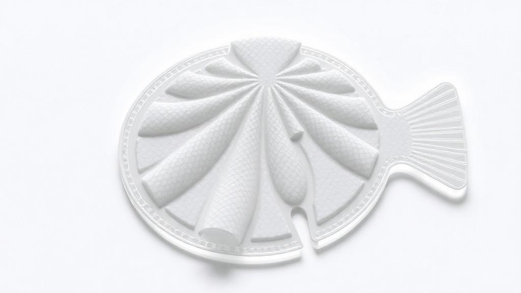
Chronic diabetic wounds are among medicine’s most stubborn adversaries. Elevated blood sugar disrupts nearly every stage of natural repair, leaving foot ulcers trapped in a state of stalled healing that can spiral into infection, hospitalization, and even amputation. Now, researchers in South Korea have engineered a 3D-printed wound patch built from two unlikely raw materials—tilapia skin and carp skin—that simultaneously calms inflammation, fights bacteria, quenches destructive oxidative stress, and coaxes new blood vessels to grow. The study, published in Materials Today Bio, demonstrates how a carefully blended fish-derived hydrogel can outperform a conventional mammalian-derived scaffold in healing diabetic wounds in mice.
The pathological environment of a diabetic wound is a tangle of interconnected failures. Excessive reactive oxygen species amplify inflammatory signaling, persistent myeloid cell activation delays the transition from inflammation to tissue proliferation, and insufficient vascularization starves the wound of the oxygen and nutrients needed for granulation and remodeling. Standard dressings offer physical coverage and moisture control but rarely intervene in these destructive feedback loops. The research team, led by investigators at Chungnam National University School of Medicine, set out to create a dressing that would actively modulate the wound microenvironment rather than simply cover it.
Decellularized extracellular matrix, or dECM, has long attracted regenerative-medicine researchers because it preserves the structural proteins, glycosaminoglycans, and other matrix-associated components that support cell adhesion, migration, and tissue remodeling. Fish skin has emerged as a particularly compelling source: it is collagen-rich, widely available as a sustainable byproduct, and sidesteps concerns tied to mammalian tissues, including pathogen transmission and cultural or religious restrictions. Clinically, acellular fish skin grafts have already shown promise for difficult-to-heal diabetic foot ulcers, lending translational weight to the approach.
Yet fish skin matrices carry a manufacturing problem. Most clinically used fish ECM materials come as sheets, grafts, or particulates with limited geometric control and poor conformal adaptation to irregular wounds. Extrusion-based 3D printing could solve this by producing patches with defined lattice geometry and interconnected porosity, but fish-derived dECM bioinks are notoriously difficult to print. Fish collagens generally have lower thermal stability than mammalian collagens, and dECM bioinks often suffer from weak mechanical integrity, batch-dependent rheology, and poor shape fidelity after extrusion. Chemical tricks like methacrylation followed by photopolymerization can help, but they add processing complexity and require light-based curing.
The Korean team’s solution was elegantly simple: blend two fish species. They prepared decellularized matrices from tilapia and carp skin using an identical workflow, then combined them at different ratios to create composite bioinks. The reasoning was that tilapia dECM, rich in collagen, could provide the structural backbone needed for thermally induced fibrillogenesis—the self-assembly of collagen molecules into a fibrillar network at body temperature—while carp dECM could contribute complementary biochemical cues, including endogenous fatty-acid-associated components. Rather than assuming that published thermal properties of isolated fish collagens would predict hydrogel behavior, the researchers determined formulation performance empirically through rheology-guided screening.
Rheological testing revealed that all formulations exhibited shear-thinning behavior, meaning viscosity drops under stress—a property ideal for extrusion through a fine nozzle. Tilapia dECM showed the highest apparent viscosity and a steep rise in storage modulus near 37 to 38 degrees Celsius, consistent with robust collagen fibrillogenesis. Carp dECM alone had much lower viscosity and only modest gelation, producing broadened, collapsed filaments when printed. But the intermediate blend, designated TdECM/CdECM-2, struck the sweet spot: continuous filaments, lattice architectures with printability indices near the ideal value of one, and no need for methacrylation or photopolymerization. After printing, the team stabilized the constructs with EDC and NHS crosslinking chemistry, preserving the lattice without light exposure.
Biochemical analysis added a fascinating layer to the story. Both decellularized matrices retained collagen, glycosaminoglycans, and elastin, but carp dECM held significantly more total free fatty acids than tilapia dECM—a difference the researchers attribute to intrinsic species composition rather than processing artifacts, since both were prepared identically. While the assay did not resolve individual fatty-acid species, the finding aligns with growing evidence that omega-3 polyunsaturated fatty acids such as EPA and DHA, which carp is known to contain, contribute antimicrobial, immunomodulatory, and pro-angiogenic activity in fish skin grafts.
In laboratory assays, the biological dividends of that compositional difference became clear. Conditioned media from carp dECM drove the strongest keratinocyte migration and endothelial tube formation, while the composite blend TdECM/CdECM-2 preserved much of that pro-migratory and pro-angiogenic activity while remaining printable. Against Staphylococcus aureus and Pseudomonas aeruginosa, the fish-derived formulations reduced bacterial survival in direct-contact tests, with carp-containing blends performing best. Live-dead staining showed compromised bacterial membrane integrity, and scanning electron microscopy revealed far fewer bacteria adhering to the composite surfaces—evidence that the effect goes beyond simple physical trapping.
The immunomodulatory results may be the most clinically significant. In macrophages stimulated to an inflammatory state, the composite scaffold suppressed expression of interleukin-6, tumor necrosis factor-alpha, and inducible nitric oxide synthase, while boosting CD206, interleukin-10, arginase 1, and transforming growth factor-beta—markers associated with pro-regenerative macrophage phenotypes. Under oxidative stress induced by hydrogen peroxide, the scaffold dramatically reduced intracellular reactive oxygen species fluorescence and upregulated the antioxidant defense genes HO-1 and Nrf2. Keratinocytes, fibroblasts, and endothelial cells grown on the printed scaffolds all showed improved metabolic activity and cytoskeletal organization under oxidative challenge.
In diabetic mice with full-thickness dorsal wounds, the printed composite patch delivered on its promise. Wounds treated with TdECM/CdECM-2 closed significantly more by day 14 than untreated or porcine-dECM-treated wounds, with histology showing more continuous epidermal coverage and markedly greater collagen deposition. Flow cytometry revealed reduced accumulation of inflammatory macrophage-enriched and eosinophil-enriched populations, along with decreased MHC-II expression on macrophages, suggesting attenuated inflammatory activation. Immunofluorescence for CD31 revealed significantly enhanced neovascularization in patch-treated wounds. Exploratory single-cell RNA sequencing further showed enriched expression of angiogenesis and repair genes, including Vegfa, Angpt1, and Hif1a, in the non-immune compartment. The authors caution that the specific contribution of carp dECM beyond tilapia alone remains unresolved, and targeted lipid profiling will be needed—but the platform stands as a striking demonstration that two supermarket fish, properly processed and printed, can yield a wound dressing that speaks the language of regenerative biology.
Subject of Research: A 3D-printed composite hydrogel wound patch made from decellularized tilapia and carp skin extracellular matrix for immunomodulatory and pro-angiogenic diabetic wound repair
Article Title: 3D-printed fish-skin-derived composite dECM hydrogel patch for immunomodulatory and pro-angiogenic diabetic wound repair
Article References: Lee, S. H., Moon, J. S., Noh, S., Yi, H.-S., & Lee, H. (2026). 3D-printed fish-skin-derived composite dECM hydrogel patch for immunomodulatory and pro-angiogenic diabetic wound repair. Materials Today Bio, 41, Article 103753. https://doi.org/10.1016/j.mtbio.2026.103753
Image Credits: AI Generated
DOI: 10.1016/j.mtbio.2026.103753
Keywords: diabetic wounds, 3D printing, decellularized extracellular matrix, fish skin, hydrogel, wound healing, immunomodulation, angiogenesis, tilapia, carp, biomaterials, oxidative stress
Cite Scienmag News
Denise Maddox. (October 16, 2026). Fish-Skin Hydrogel Patch 3D-Printed to Heal Stubborn Diabetic Wounds. Scienmag. https://scienmag.com/fish-skin-hydrogel-patch-3d-printed-to-heal-stubborn-diabetic-wounds/
Denise Maddox. "Fish-Skin Hydrogel Patch 3D-Printed to Heal Stubborn Diabetic Wounds." Scienmag, 16 October 2026, https://scienmag.com/fish-skin-hydrogel-patch-3d-printed-to-heal-stubborn-diabetic-wounds/. Accessed 16 October 2026.
Denise Maddox. "Fish-Skin Hydrogel Patch 3D-Printed to Heal Stubborn Diabetic Wounds." Scienmag. October 16, 2026. https://scienmag.com/fish-skin-hydrogel-patch-3d-printed-to-heal-stubborn-diabetic-wounds/


