


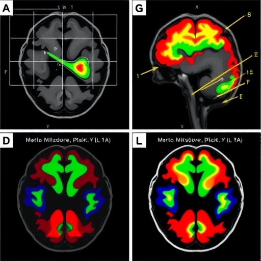
A new study has brought scientists closer to observing how inflammation during pregnancy may alter the earliest stages of human brain development. Using three-dimensional tissue models made from human fetal brain samples, researchers modeled maternal immune activation and identified the signaling molecule interleukin-17A, or IL-17A, as a major driver of abnormal cortical development. The findings, published in Nature Neuroscience, offer a human-based experimental system for investigating how immune signals outside the brain can influence the formation of neural circuits before birth.
Maternal immune activation refers to the biological response triggered when a pregnant individual experiences infection or significant inflammation. Epidemiological studies have associated certain prenatal inflammatory exposures with an increased likelihood of neurodevelopmental conditions, including autism spectrum disorder, schizophrenia, and intellectual disability. Yet the mechanisms behind these associations have remained difficult to study. Animal models have provided important clues, but differences between species can limit their ability to reproduce the timing, cellular organization, and molecular environment of the developing human cortex.
The researchers addressed this challenge with “brain cerebroids,” three-dimensional, ex vivo models that preserve features of developing human fetal brain tissue. Unlike simplified cultures composed of a single cell type, these structures retain a more complex arrangement of neural progenitors, immature neurons, and supporting cells. Their three-dimensional architecture allows cells to communicate and organize in ways that more closely resemble the fetal cortex, including the production and movement of neurons that ultimately establish the layered structure of the brain’s outer region.
To recreate aspects of maternal immune activation, the team exposed the fetal brain cerebroids to inflammatory conditions associated with immune signaling during pregnancy. The central focus was IL-17A, a cytokine produced primarily by activated immune cells, particularly a subset of T cells known as T helper 17 cells. Cytokines are small signaling proteins that coordinate immune responses, but they can also affect tissues beyond the immune system. In the developing brain, inappropriate or excessive exposure to such signals may influence cell proliferation, migration, differentiation, and the formation of connections between neurons.
The study found that IL-17A exposure was linked to disruptions in cortical development within the human cerebroids. These effects are described as “IL-17A-driven,” indicating that the cytokine was not merely associated with the developmental changes but played a functionally important role in producing them. The disturbances affected the organization and behavior of developing neural cells, processes that are essential for building the cortex. During normal development, neural progenitor cells generate new neurons in tightly regulated sequences, while young neurons migrate to their appropriate positions. Interference with either process can alter the structure and connectivity of later-forming brain tissue.
The results are especially significant because they were observed in human fetal brain material rather than inferred only from animal studies. Previous research in mice has implicated maternal immune activation and IL-17A in changes to cortical organization and behavior in offspring. However, the human cerebroid model provides an opportunity to examine whether similar immune mechanisms operate in human tissue, where the timing of neurogenesis, cell-type composition, and cortical architecture differ from those of rodents. The approach may also help reveal human-specific responses that conventional laboratory animals cannot capture.
The findings do not mean that IL-17A exposure during pregnancy inevitably causes a neurodevelopmental condition, nor do they establish that prenatal inflammation is a single explanation for such conditions. Brain development is shaped by a combination of genetic factors, immune signals, environmental influences, and developmental timing. The experiments also took place in an ex vivo model, outside the full physiological environment of a living pregnancy. Cerebroids cannot reproduce the placenta, maternal circulation, hormonal changes, or the complete immune system, all of which influence how inflammatory signals reach the fetus and how long they remain active.
Nevertheless, the model could become a powerful platform for testing mechanisms and potential interventions. Researchers may use it to determine how IL-17A changes gene activity in neural progenitors, whether it alters communication between neurons and glial cells, and which molecular pathways make some developing cells more vulnerable than others. Because the tissue is human and three-dimensional, it may also support studies of patient-specific genetic risk, allowing investigators to compare how different backgrounds respond to the same inflammatory signal.
The work also highlights a broader shift in neuroscience toward experimental systems that combine human tissue with controlled laboratory manipulation. Understanding prenatal inflammation is difficult in clinical studies because immune exposure cannot be ethically assigned or precisely timed in pregnant people. Brain cerebroids offer a way to isolate individual components of that complex biology while preserving more of the developing tissue’s natural organization than conventional two-dimensional cell cultures. As these models improve, they may help distinguish temporary developmental changes from persistent alterations in cortical circuitry and identify when immune signaling is most consequential.
By showing that IL-17A can disrupt human cortical development in a three-dimensional ex vivo system, the study gives researchers a clearer biological target in the search for links between prenatal inflammation and later neurodevelopment. Its most immediate contribution is not a new diagnosis or treatment, but a more precise experimental framework: one that connects maternal immune signaling to cellular events in the developing human cortex. That connection could guide future work on prevention, therapeutic timing, and the complex origins of developmental differences long before the first symptoms become visible.
Cite Scienmag News
Cassandra Pierce. (August 11, 2026). Fetal brain models show IL-17A disrupts cortical development after maternal immune activation. Scienmag. https://scienmag.com/fetal-brain-models-show-il-17a-disrupts-cortical-development-after-maternal-immune-activation/
Cassandra Pierce. "Fetal brain models show IL-17A disrupts cortical development after maternal immune activation." Scienmag, 11 August 2026, https://scienmag.com/fetal-brain-models-show-il-17a-disrupts-cortical-development-after-maternal-immune-activation/. Accessed 4 September 2026.
Cassandra Pierce. "Fetal brain models show IL-17A disrupts cortical development after maternal immune activation." Scienmag. August 11, 2026. https://scienmag.com/fetal-brain-models-show-il-17a-disrupts-cortical-development-after-maternal-immune-activation/


