


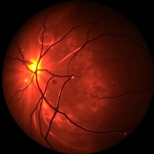
In a groundbreaking study, researchers have developed a multi-stage learning approach aimed at revolutionizing the visualization of microcystic macular edema (MME) using Optical Coherence Tomography (OCT) images. This condition, characterized by the accumulation of fluid in the retina, is particularly challenging to diagnose and monitor. The researchers, led by Vidal et al., have created a new computational method that enhances the clarity and interpretability of OCT images, making it easier for healthcare providers to recognize and assess the severity of MME in their patients.
The core of this research lies in the use of advanced machine learning algorithms designed specifically for image processing. Machine learning has proven to be a powerful tool in various medical applications, and this study leverages its potential by incorporating a multi-stage learning framework. Each stage of the algorithm is finely tuned to focus on different aspects of the OCT images, allowing for a more nuanced understanding of the changes occurring within the retina due to MME.
One of the standout features of this multi-stage learning system is its capability to improve upon traditional image processing methods. Conventional techniques often struggle to isolate specific features within OCT images, leading to potential diagnostic inaccuracies. In contrast, the researchers’ approach employs a sequence of preprocessing, feature extraction, and classification steps. This systematic methodology ensures that the final visualization presented to clinicians is not only high in fidelity but also rich in clinically relevant information.
An essential component of their technique involves a deep learning architecture that utilizes convolutional neural networks (CNNs). CNNs have become the gold standard in image analysis due to their ability to learn spatial hierarchies of features. By training these networks on a diverse dataset of OCT images, the researchers were able to create a model that significantly enhances the detection and visualization of microcystic changes associated with MME.
The implications of this research extend beyond the immediate enhancement of OCT image visualization. By improving the diagnostic capability for MME, healthcare providers can make more informed decisions regarding treatment options for patients. The early detection and clear visualization of microcystic changes can lead to timely interventions, potentially modifying the disease’s progression and preserving visual function.
In addition to the technical advancements, the researchers have made significant strides in making their findings accessible to clinicians. They understand that cutting-edge technology must be user-friendly to be effectively integrated into clinical practice. As a result, the team has focused on creating intuitive visualization tools that can easily be operated within existing healthcare frameworks, facilitating seamless integration into everyday use.
Furthermore, the study highlights the importance of collaborative efforts in medical research. The interprofessional team comprising engineers, medical professionals, and data scientists enabled a comprehensive approach to the challenges posed by MME. By pooling their diverse skill sets, they were able to address both the technical and clinical dimensions of the problem, paving the way for future innovations in OCT imaging and beyond.
Another notable aspect of this research is its potential to influence education and training for ophthalmologists and other medical professionals. As new technologies emerge, continuous education is vital to ensure that healthcare providers are familiar with these tools’ capabilities and limitations. By disseminating their findings through publications, workshops, and seminars, the researchers aim to empower the next generation of clinicians with the knowledge necessary to utilize advanced imaging techniques effectively.
To ensure the robustness of their findings, the researchers conducted extensive validation and testing of their model. They compared their novel approach against several existing methods, demonstrating superior performance in terms of accuracy and interpretability. These findings underline the potential for their multi-stage learning technique to set a new standard in OCT imaging, highlighting the need for ongoing refinement and testing in diverse clinical settings.
Moreover, this study contributes to the growing body of evidence that machine learning can significantly enhance diagnostic imaging. As the field of medical imaging continues to evolve, there will likely be more emphasis on the development of AI-assisted tools that can provide real-time analysis and feedback to clinicians during examinations. The researchers believe that their approach could be a steppingstone toward more comprehensive AI models that incorporate patient history and other relevant factors to deliver tailored diagnostic insights.
As the healthcare industry increasingly embraces technological advancements, the cooperation between physicians and data scientists will be crucial in leveraging these innovations. This partnership will be essential for translating complex data into actionable clinical decisions, ultimately benefiting patient care.
In conclusion, the multi-stage learning framework developed by Vidal et al. presents a promising advancement in the visualization of microcystic macular edema in OCT images. By combining sophisticated machine learning techniques with user-friendly design, this research provides a blueprint for the future of medical imaging. Its implications could resonate far beyond MME, offering insights and capabilities that could transform various fields within healthcare, making diagnostic processes smarter, faster, and more efficient.
The journey towards revolutionizing optical imaging through this study is only the beginning. As research progresses, the integration of more powerful computational tools and databases will likely yield even more sophisticated imaging modalities, enhancing patient outcomes in the realm of ophthalmology and other medical specialties.
Cite Scienmag News
Ophelia Keating. (August 29, 2025). Enhanced Visualization of Microcystic Macular Edema in OCT. Scienmag. https://scienmag.com/enhanced-visualization-of-microcystic-macular-edema-in-oct/
Ophelia Keating. "Enhanced Visualization of Microcystic Macular Edema in OCT." Scienmag, 29 August 2025, https://scienmag.com/enhanced-visualization-of-microcystic-macular-edema-in-oct/. Accessed 4 September 2026.
Ophelia Keating. "Enhanced Visualization of Microcystic Macular Edema in OCT." Scienmag. August 29, 2025. https://scienmag.com/enhanced-visualization-of-microcystic-macular-edema-in-oct/


