


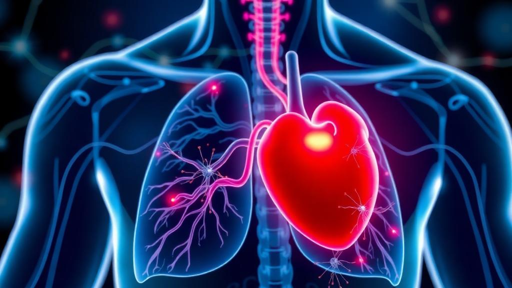
When the heart is temporarily deprived of blood during cardiac surgery and then flooded with oxygen-rich flow again, the damage does not stay confined to the chest. The lungs, exquisitely sensitive to the inflammatory storm that follows ischemia and reperfusion, often bear a heavy share of the injury. Major pulmonary complications occur in roughly 19 to 40 percent of patients undergoing cardiac surgery, a striking contrast to the 5 to 8 percent seen after non-cardiac procedures, and they translate into longer hospital stays, higher costs, and setbacks for enhanced recovery after surgery programs. A new study published in Physiological Reports now offers a surprising candidate for protection: electroacupuncture, delivered days before the insult, appears to blunt lung injury by suppressing a well-known inflammatory signaling axis.
The research team, working at the Laboratory Animal Center of Shanghai University of Traditional Chinese Medicine, set out to answer two linked questions. First, could they establish a stable and reproducible rat model of lung injury driven by myocardial ischemia and reperfusion? Second, could pretreatment with electroacupuncture reduce that injury, and if so, did the HMGB1/RAGE pathway, a pair of molecules long implicated in acute lung inflammation, sit at the center of the mechanism? Their answer to both questions, based on a series of five carefully controlled experiments in male Sprague-Dawley rats, was yes.
Building the model required calibrating the injury. The researchers ligated the left anterior descending coronary artery in anesthetized rats for 30 minutes, 45 minutes, or one hour, then released the snare to allow 24 hours of reperfusion, confirming ischemia by visible cyanosis of the ventricular wall and ST-segment elevation on the electrocardiogram. Only the one-hour ischemia protocol produced consistent, measurable lung damage. In those animals, arterial oxygen partial pressure and the oxygenation index fell significantly, the alveolar-arterial oxygen gradient rose, and the lung wet-to-dry weight ratio, a standard gauge of pulmonary edema, climbed sharply. Under the microscope, the lungs showed alveolar collapse, hemorrhage, thickened septa, and dense inflammatory infiltration, and immunostaining revealed abundant cleaved caspase-3, a hallmark of apoptosis. Shorter ischemic periods produced weaker or statistically insignificant changes, establishing one hour of ischemia followed by 24 hours of reperfusion as the reliable model.
With the model in hand, the team turned to electroacupuncture. They stimulated two classical acupoints, Zusanli (ST36) on the hind limb and Feishu (BL13) on the back near the lungs, using a 2/20 Hz dense-disperse waveform for 30 minutes per session. The critical variable was timing. A single 30-minute session immediately before ischemia did essentially nothing: blood gas values, edema measures, injury scores, and apoptotic cell counts in that group were indistinguishable from the untreated ischemia-reperfusion group. But when the same stimulation was repeated over three consecutive days before the procedure, the picture changed dramatically. Oxygenation improved, the wet-to-dry ratio dropped, lung injury scores fell, and cleaved caspase-3 staining diminished significantly compared with injured animals that received no pretreatment.
The protective effect was accompanied by a clear anti-inflammatory signature. Quantitative PCR showed that ischemia-reperfusion drove up the messenger RNA of the pro-inflammatory cytokines IL-1β and IL-6 and the chemokines CXCL1 and CXCL2 in lung tissue. Three days of electroacupuncture pretreatment significantly reduced all four transcripts at four hours of reperfusion, with IL-6 and CXCL1 still suppressed at 24 hours. Immunohistochemistry told a parallel story at the cellular level: myeloperoxidase-positive neutrophils and F4/80-positive macrophages, the two dominant infiltrating populations, flooded the injured lungs after reperfusion but were markedly fewer in the pretreated animals at both early and late time points.
The molecular centerpiece of the study was HMGB1, a nuclear non-histone protein that escapes from stressed cells and acts as a danger signal when it binds transmembrane receptors, most notably RAGE and the Toll-like receptors. Western blotting and PCR revealed that ischemia-reperfusion significantly increased both HMGB1 and RAGE in lung tissue, and electroacupuncture pretreatment brought both back down at four and 24 hours of reperfusion. Because HMGB1 is known to recruit and activate inflammatory cells in many forms of acute lung injury, the authors reasoned that this axis could be the plausible bridge between the systemic inflammatory surge triggered by cardiac ischemia and the pulmonary damage that follows.
A key question was where the lung HMGB1 actually came from. The injured heart itself is an obvious suspect. Yet when the researchers measured HMGB1 expression in heart tissue, they found it elevated only in the earliest phase of inflammation and essentially normal by 24 hours of reperfusion, precisely when lung HMGB1 remained high. Moreover, electroacupuncture reduced lung HMGB1 without changing myocardial infarct size, measured by triphenyltetrazolium chloride staining, indicating that the treatment was not simply shrinking the cardiac wound and thereby sending fewer danger signals. The accumulating HMGB1 in the lungs, the data suggested, was generated locally rather than imported from the heart.
To test causality more directly, the team delivered recombinant HMGB1 protein directly into the lungs via intubation-mediated intratracheal administration before reperfusion. Adding the protein to injured animals worsened respiratory dysfunction, edema, and histological injury to some degree. More tellingly, in rats that had received the three-day electroacupuncture pretreatment, intratracheal recombinant HMGB1 significantly raised lung HMGB1 and RAGE expression, lowered oxygenation indices, increased the wet-to-dry ratio, worsened injury scores, and boosted apoptotic cell counts. In other words, flooding the lungs with the very molecule the therapy suppresses partially reversed its benefits, evidence that HMGB1 is not merely a bystander but a functional target of the electroacupuncture effect.
The authors are candid about the limits of their work. The validation relied on recombinant protein challenge rather than genetic knockout, which leaves some causal uncertainty, and the sample sizes were small, with five rats per group. All experiments were conducted in male animals, and no clinical data exist yet to confirm that the findings translate to patients awaiting cardiac surgery. Larger, multicenter human studies will be needed before electroacupuncture pretreatment can be recommended as a standard element of perioperative lung protection.
Even so, the study adds a compelling piece to a growing body of evidence that neuromodulation through acupuncture-like stimulation can precondition distant organs against surgical stress. Previous work has shown electroacupuncture protecting the lungs during cardiopulmonary bypass by dampening inflammasome activation and oxidative stress pathways, and the new findings extend that logic to the specific HMGB1/RAGE axis in the setting of myocardial ischemia and reperfusion. If the mechanism holds in humans, a low-side-effect intervention delivered in the days before surgery could help reduce the burden of postoperative pulmonary complications, aligning with the principles of enhanced recovery after surgery and broadening the toolkit of perioperative organ protection.
Subject of Research: Electroacupuncture pretreatment and protection against myocardial ischemia/reperfusion-induced lung injury via the HMGB1/RAGE pathway in rats
Article Title: Electroacupuncture pretreatment alleviates myocardial ischemia/reperfusion‐induced lung injury by inhibiting the expression of HMGB1/RAGE in male rats
Article References: Xie, C., Zhang, J., Zhang, Y., Chi, H., Yong, Y., & Song, J. (2026). Electroacupuncture pretreatment alleviates myocardial ischemia/reperfusion‐induced lung injury by inhibiting the expression of HMGB1 / RAGE in male rats. Physiological Reports, 14(18), Article e71066. https://doi.org/10.14814/phy2.71066
Image Credits: AI Generated
DOI: 10.14814/phy2.71066
Keywords: electroacupuncture, myocardial ischemia-reperfusion, lung injury, HMGB1, RAGE, inflammation, acute lung injury, cardiac surgery, pulmonary complications, rat model, preconditioning, enhanced recovery after surgery
Cite Scienmag News
Ophelia Keating. (September 30, 2026). Electroacupuncture Before Heart Surgery Shields the Lungs by Taming a Key Inflammatory Protein. Scienmag. https://scienmag.com/electroacupuncture-before-heart-surgery-shields-the-lungs-by-taming-a-key-inflammatory-protein/
Ophelia Keating. "Electroacupuncture Before Heart Surgery Shields the Lungs by Taming a Key Inflammatory Protein." Scienmag, 30 September 2026, https://scienmag.com/electroacupuncture-before-heart-surgery-shields-the-lungs-by-taming-a-key-inflammatory-protein/. Accessed 4 October 2026.
Ophelia Keating. "Electroacupuncture Before Heart Surgery Shields the Lungs by Taming a Key Inflammatory Protein." Scienmag. September 30, 2026. https://scienmag.com/electroacupuncture-before-heart-surgery-shields-the-lungs-by-taming-a-key-inflammatory-protein/


