


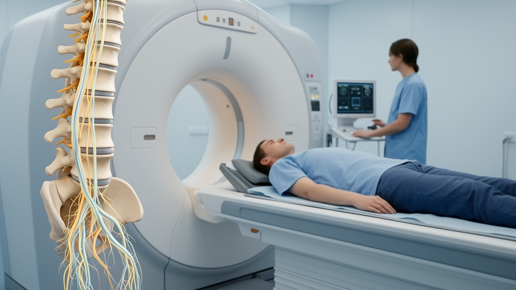
Diffusion tensor imaging, an MRI technique that maps the directional movement of water molecules, showed potential for tracking recovery in compressed lumbar nerve roots, predicting postoperative outcomes and assessing whether conservative treatment reduces nerve injury. A 2026 narrative review of research in lumbar disc herniation and lumbar spinal stenosis concluded that the technique can reveal microstructural changes not readily characterized by conventional MRI, but warned that most findings remain preliminary and no imaging threshold is ready for routine clinical decisions.
The review focused on two forms of prevention. Primary prevention concerns identifying warning signs before an injury or disabling recurrence occurs, while secondary prevention concerns evaluating rehabilitation to reduce long-term disability. DTI may contribute to both by measuring changes in nerve roots and paraspinal muscles, structures that support the lumbar spine. The authors identified potential applications in postoperative monitoring, preoperative prognosis, conservative treatment assessment, paraspinal muscle evaluation and military training, while emphasizing that evidence specific to military personnel is especially limited.
DTI applies diffusion-sensitive magnetic-field gradients in at least six directions to measure how freely water moves through tissue. In nerve tissue, water generally diffuses more readily along the long axis of axons than across them, a property known as anisotropy. The most widely studied DTI measure, fractional anisotropy, or FA, ranges from 0 to 1: higher values indicate more directional diffusion and orderly fiber alignment, whereas lower values are consistent with disrupted or disorganized fibers. Other measures include mean diffusivity, axial diffusivity and radial diffusivity, which describe overall, lengthwise and crosswise diffusion, respectively. Together, these metrics can provide information about axonal injury, demyelination, edema and tissue organization, although DTI does not directly establish the cause of a measured change.
Studies in patients with nerve-root compression support the biological relevance of these measurements. In a small study of 20 elderly patients with L5 compression caused by spinal canal or foraminal stenosis, Hughes and colleagues found lower FA and higher mean, axial and radial diffusivity in the compressed L5 root than in uncompressed L4 or S1 roots. The pattern was similar whether compression arose from stenosis or a herniated disc. Another prospective physical-therapy study of 14 patients with chronic unilateral lumbosacral radiculopathy found that FA in the affected root rose from 0.226 to 0.268 and apparent diffusion coefficient fell from 1.764 × 10⁻³ to 1.616 × 10⁻³ mm²/s after six weeks of multimodal therapy. Pain and disability scores improved beyond their reported minimum clinically important differences, linking imaging change with recovery, but the study had no control group and did not report the full DTI parameter set.
Postoperative evidence suggests that DTI may also reveal gradual neural recovery. In a prospective longitudinal study of 19 patients with lumbar disc herniation who underwent percutaneous transforaminal endoscopic discectomy, affected-side FA remained below the healthy-side value 30 days after surgery but showed no significant difference by 90 days. For the L5 roots, for example, mean FA was 0.354 before surgery, 0.408 at 30 days and 0.421 at 90 days, compared with 0.442 on the healthy side; the S1 roots followed a similar trajectory. A separate longitudinal series of seven patients with lumbar spinal stenosis found no significant change in overall FA after interspinous process device implantation, yet FA increased within the foramen and other diffusion measures decreased in its anterior segment as pain improved. These findings indicate that regional analysis may reveal changes that a whole-segment average can obscure.
Preoperative DTI measurements have also been associated with later outcomes. In a prospective single-centre study of 117 patients with lumbar disc herniation, Wu and colleagues reported that affected-side FA was strongly associated with favorable surgical outcomes, with an area under the receiver operating characteristic curve of 0.900 and a 95% confidence interval of 0.829–0.971. A proposed FA cutoff above 0.212 had sensitivity of 0.99 and specificity of 0.77, but the threshold has not been externally validated. Other findings are similarly promising but not yet definitive: a retrospective analysis of 364 patients found that lower FA and higher apparent diffusion coefficient independently predicted poor conservative-treatment outcomes, while a prospective study of 33 patients with stenosis reported an FA below 0.295 as predictive of poor prognosis. Importantly, those thresholds are not interchangeable and show overlap between outcome groups, so they should not currently guide treatment choices.
DTI may also assist surgical planning. In a retrospective case-control study of 128 patients undergoing extreme lateral interbody fusion, investigators used three-dimensional reconstructions of the psoas major and nerve pathways to select personalized entry points. Compared with a conventional approach, DTI-guided planning was associated with fewer immediate postoperative complications, including thigh pain or numbness in two rather than ten patients and lower-limb motor dysfunction in one rather than eight. This is an association within a planned comparison, not proof that imaging itself prevented the complications, and the approach requires prospective validation across surgical techniques and spinal levels.
Beyond nerve roots, DTI can assess the paraspinal muscles that stabilize the lumbar spine. Muscle fibers also conduct water preferentially along their long axes, allowing FA to reflect fiber orientation and fascicular organization. Studies have found lower FA and higher apparent diffusion coefficient in people with persistent low back pain, with the measures associated with pain severity and fatty infiltration. Other research detected age-related differences that conventional cross-sectional area or fat-fraction measures might not reveal. Animal experiments have shown that FA changes accompany a shift from type I to type II muscle fibers and functional deterioration, providing complementary mechanistic evidence rather than direct proof of clinical benefit in people.
The military application remains the most exploratory part of the research framework. High-intensity training places repeated loads on the lumbar paraspinal muscles, and low back pain is common among service personnel. In a cross-sectional study of active-duty Marines positioned in standing, weight-bearing, sitting and prone-with-elbow-support postures, erector-spinae FA predicted lumbar lordosis, lumbosacral extension and sacral tilt. Lower FA was inversely associated with muscle-fiber size and correlated with poorer lumbar stability. The design cannot establish causation or demonstrate that DTI predicts future injury, and direct military evidence is scarce. Proposed uses such as quantifying training load, warning of muscle injury and assessing readiness to resume demanding activity therefore remain research questions, not established safety procedures.
Across the literature, the central obstacle is not a lack of biological plausibility but a lack of standardization and validation. FA and diffusion measurements can vary with scanner field strength, diffusion-encoding settings, echo time, region-of-interest placement, tractography methods and artifact correction. Most studies have small samples, short follow-up or no control group, and the review rates the key evidence as low or moderate rather than high. Future work needs large, multicenter prospective cohorts, consistent acquisition protocols, external testing of thresholds and longer follow-up, particularly among military personnel. Until then, DTI is best regarded as a research tool and potential adjunct to conventional assessment, not a stand-alone guide to surgery, rehabilitation or return to duty.
Subject of Research: Diffusion tensor imaging for rehabilitation and prevention of lumbar degenerative diseases
Article Title: Diffusion tensor imaging for rehabilitation and prevention of lumbar degenerative diseases: a narrative review
Article References: Zhang, N., Zhang, B., Zhao, Z., Xu, D., & Xu, Y. (2026). Diffusion tensor imaging for rehabilitation and prevention of lumbar degenerative diseases: a narrative review. Frontiers in Medicine, 13, Article 1959293. https://doi.org/10.3389/fmed.2026.1959293
Image Credits: AI Generated
DOI: 10.3389/fmed.2026.1959293
Keywords: diffusion tensor imaging, lumbar disc herniation, lumbar spinal stenosis, nerve-root recovery, low back pain, rehabilitation imaging, paraspinal muscles, fractional anisotropy, military training, clinical validation, Diffusion, tensor
Cite Scienmag News
Ophelia Keating. (October 25, 2026). Diffusion imaging tracks nerve recovery after lumbar surgery. Scienmag. https://scienmag.com/diffusion-imaging-tracks-nerve-recovery-after-lumbar-surgery/
Ophelia Keating. "Diffusion imaging tracks nerve recovery after lumbar surgery." Scienmag, 25 October 2026, https://scienmag.com/diffusion-imaging-tracks-nerve-recovery-after-lumbar-surgery/. Accessed 25 October 2026.
Ophelia Keating. "Diffusion imaging tracks nerve recovery after lumbar surgery." Scienmag. October 25, 2026. https://scienmag.com/diffusion-imaging-tracks-nerve-recovery-after-lumbar-surgery/


