In a groundbreaking study published in the prestigious journal Nature, researchers have uncovered a previously unrecognized early developmental organization in the peripheral nervous system of humans. Spearheaded by Xiaoxu Yang, Ph.D., at University of Utah Health, along with Keng Ioi Vong, Ph.D., and Joseph Gleeson, M.D., at the University of California San Diego, this multidisciplinary team employed innovative genetic lineage tracing techniques to rewrite a fundamental principle of developmental biology. Their investigation conclusively demonstrates that sensory and sympathetic ganglia—the complex nerve clusters integral to sensory perception and autonomic function—originate from distinct precursor cell populations well before these cells migrate from the neural tube, challenging the long-standing dogma of neural crest cell fate determination.
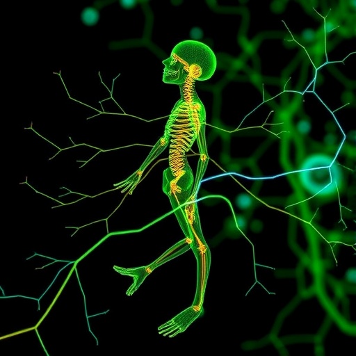
The peripheral nervous system connects the brain to organs, muscles, and skin, enabling sensations and involuntary responses vital for survival. It develops remarkably early in the embryo, beginning with neural crest cells, which are multipotent progenitors first emerging from the neural tube, the embryonic structure that eventually forms the central nervous system. For decades, the accepted theory posited that these neural crest cells, once they delaminate and migrate away from the neural tube, subsequently differentiate into various ganglionic lineages. However, the new findings reveal that lineage specification is preordained even before migration, indicating a sophisticated pre-patterning within the neural tube.
The key to this discovery lies in an approach leveraging the subtle mosaicism of the human genome accrued over a lifetime. Each cell’s DNA is not a perfect replica due to random mutations arising during cell division in embryogenesis. These somatic mutations serve as natural barcodes, enabling scientists to reconstruct the developmental lineage tree of cells—a feat previously elusive, especially in humans due to ethical and technical constraints. By isolating adult cells from sensory and sympathetic ganglia and sequencing their genomes with exceptional precision, the researchers traced back the shared mutation signatures to map cell lineage trajectories.
This method illuminated that the progenitor populations destined for sensory or sympathetic ganglia are genetically distinct groups within the neural tube rather than a homogenous pool of migratory cells differentiating later. Such delineation of fates implies an intrinsic programming at the earliest stages, where environmental cues and gene regulatory networks likely prime these cells towards unique identities. The team’s complementary experiments in animal models like mice and quail corroborated these findings, revealing that the migration path post-delamination follows a highly regulated pattern orchestrated by molecular signals guiding each neural crest subset to its eventual anatomical locale.
Crucially, this early commitment highlights how developmental disorders originating from neural crest derivatives might arise due to disruptions affecting these primordial populations or their initial specification. The peripheral nervous system’s architecture stems from this precise choreography; deviations may underlie congenital conditions involving sensory deficits or autonomic dysfunctions. Furthermore, childhood cancers such as neuroblastoma and neurofibromatosis, both linked to aberrant neural crest cell development, might be better understood and therapeutically targeted by considering cell fate decisions made within the neural tube itself, well before noticeable phenotypes emerge.
The implications extend beyond pathogenesis to preventative health strategies, underscoring the critical nature of the earliest embryonic environment. Yang and colleagues emphasize the importance of folic acid supplementation prior to and during early pregnancy, a practice already known to reduce neural tube defects, as the neural crest cells’ formation and differentiation are intensely susceptible during these initial stages. This intersection of molecular lineage tracing data and clinical recommendations offers renewed insight into how maternal health directly influences intricate developmental processes.
By uncovering this paradigm shift, the study not only advances developmental neuroscience but also exemplifies the power of genomic technologies to backtrack cell history. The mosaic barcode approach opens avenues to explore other human-specific developmental timelines previously inaccessible through conventional model organisms or embryological observation. It also poses profound questions about the molecular mechanisms enforcing early cell identity segregation and how these mechanisms integrate spatial and temporal developmental cues.
Moreover, detailing the distinct origin and migration paths of sensory and sympathetic ganglia provides a refined anatomical and functional framework. Sensory ganglia process external stimuli—such as touch, pain, and smell—feeding information into central processing centers, while sympathetic ganglia regulate involuntary physiological activities, including heart rate and respiration. Understanding their exact developmental origins enables researchers to pinpoint the genesis of neural circuitries underpinning these diverse yet essential biological functions.
This new knowledge contributes to a holistic understanding of how the peripheral nervous system is meticulously assembled from cellular subsets predetermined for specialized roles. It suggests that future regenerative medicine approaches may harness these early lineage commitments to engineer precise cell types for transplantation or repair. By manipulating the molecular determinants responsible for early cell fate decisions within the neural tube, therapies could achieve more effective restoration of function in neurodegenerative diseases or injury.
The collaborative effort reflects a synthesis of developmental biology, genomics, and imaging technologies, supported by a range of institutions and funding bodies including the National Institutes of Health, Simons Foundation, and specialized stem cell research programs. This integrative research model exemplifies the cutting-edge science needed to unravel the complex origins of human biology and disease.
In conclusion, this study revolutionizes our perception of neural crest cell differentiation and peripheral nervous system development. It demonstrates that nerve clusters’ cellular destiny is carved within the neural tube itself during the earliest embryonic stages, preceding migration and differentiation. By deploying innovative barcode lineage tracing, the researchers have charted a more intricate and informative developmental map. This knowledge heralds new pathways to investigate congenital neurological disorders and advance clinical interventions aimed at children affected by conditions rooted in peripheral nervous system malformations.
Subject of Research: Animals
Article Title: Developmental organization of sensory and sympathetic ganglia
News Publication Date: 1-Apr-2026
Web References:
Developmental Organization of Sensory and Sympathetic Ganglia – Nature
Video summary of the study
Image Credits: Melanie White, DPhil, University of Queensland
Keywords: Developmental biology; Developmental neuroscience; Neural crest; Peripheral nervous system; Sensory neurons
