


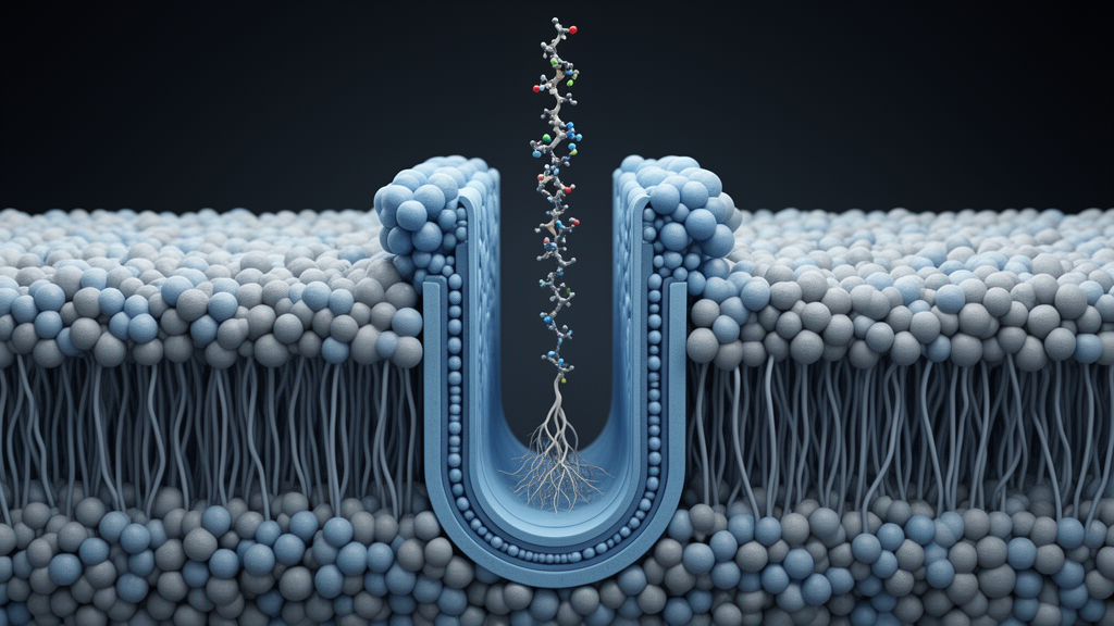
Gram-negative bacteria are among the most formidable pathogens in modern medicine, and a large part of their resilience comes from an architectural feature: an outer membrane that is studded on its outer face with lipopolysaccharide, or LPS, a molecule whose tightly packed, negatively charged structure excludes many otherwise effective antibiotics. Building and maintaining this barrier is a monumental logistics problem. Every time a Gram-negative cell divides, millions of LPS molecules must be ferried from the inner membrane, where they are synthesized, across the aqueous periplasm, and into the outer leaflet of the outer membrane. A dedicated molecular machine called the Lpt system performs this task, and a new study published in Nature has now captured, in unprecedented structural detail, how this machine decides when to work and when to idle.
The Lpt machinery is not a single membrane protein but a trans-envelope assembly that spans the entire cell envelope. At the inner membrane sits an ATP-binding cassette transporter complex known as LptB2FGC, which extracts LPS from the inner leaflet of the inner membrane. At the outer membrane, a translocon called LptDE receives the LPS and inserts it into the outer leaflet. Connecting these two membrane-embedded endpoints is LptA, a soluble periplasmic protein that assembles with both membrane complexes to form a continuous protein bridge stretching roughly 280 angstroms from membrane to membrane. Energy from ATP binding and hydrolysis in the cytoplasm drives LPS along this bridge, against its concentration gradient, all the way to the cell surface.
What has long puzzled researchers is whether this transport process is regulated. Cells must prevent toxic accumulation of LPS in the inner membrane, and one known safeguard is to shut down LPS biosynthesis when levels rise too high. But whether the transport machinery itself modulates its activity in response to substrate availability remained unclear. The new work, led by Rebecca J. Taylor, Karanbir S. Pahil, Alessio Caruso, Bailey Plaman, Stephen A. Early and Sebastian J. Rowe in Daniel Kahne’s laboratory at Harvard University, together with colleagues at Ohio State University and Harvard Medical School, answers this question by combining cryo-electron microscopy with a battery of biochemical and genetic experiments.
Capturing the intact bridge was itself a technical tour de force. Assembled bridges dissociate during purification, so the team reconstituted the complex in vitro from separately purified subcomplexes. They used a fusion of LptC and LptA, joined by a flexible poly-glycine-serine linker, which had previously been shown to support bridge formation and to be more stable than the wild-type arrangement. To lock the bridge together for imaging, they incorporated the photocrosslinkable amino acid p-benzoyl-L-phenylalanine at strategic positions in both LptD and the LptC-LptA fusion, then covalently stitched the two membrane complexes together with ultraviolet light. The resulting dumbbell-shaped particles, imaged on a Titan Krios microscope, yielded local refinements of the inner-membrane and outer-membrane portions at approximately 3.3 and 3.4 angstroms respectively.
The resulting structures revealed a striking conformational switch. In the LPS-bound state of the full bridge, clear density for an LPS molecule was visible in the lumen formed by the transmembrane helices of LptF and LptG, but the transmembrane helix of LptC, known as TM-LptC, was absent from the density map, indicating that it had become disordered or displaced. In the LPS-free bridge, the situation reversed: TM-LptC was well resolved in its resting position and no LPS occupied the lumen. Critically, a structure of the partial bridge, LptB2FGCA, with LPS bound in the lumen showed both the lipid substrate and an ordered TM-LptC. This comparison demonstrated that TM-LptC dissociation requires not merely the presence of LPS, but the assembly of the complete trans-envelope bridge.
The mechanism underlying this switch involves a subtle rearrangement of the LptG transmembrane helices. When the full bridge is loaded with LPS, helices 1 through 3 of LptG shift from a more open conformation to a constricted one, forming stabilizing contacts with the bound LPS molecule. This constriction physically displaces TM-LptC from its binding site. The displacement has functional consequences: in scintillation proximity assays using a catalytically inactive LptB variant, the full bridge bound ATP with an equilibrium dissociation constant of roughly 20 micromolar when LPS was present, matching the high affinity of the LptB2FG complex lacking TM-LptC, whereas the LPS-free bridge bound ATP with a lower affinity of about 40 micromolar, matching that of the intact LptB2FGC transporter. In other words, loading the bridge with its substrate converts it into a primed, high-affinity state.
ATPase measurements reinforced the picture. Proteoliposomes containing the inner-membrane complex with the LptC-LptA fusion showed little change in ATP hydrolysis when LptDE was added, unless LPS was also present, in which case ATPase activity increased markedly as more LptDE-containing proteoliposomes were supplied. This finding resolves a long-standing puzzle: unlike typical ABC transporters, the isolated LptB2FGC complex shows no substrate-stimulated ATPase activity. The activation is not a property of the inner-membrane complex alone but emerges only when the complete bridge is assembled and loaded with LPS, coupling transport activity to both bridge formation and substrate availability.
In vivo experiments confirmed that this conformational switch is not an artifact of in vitro reconstitution. A mutation in LptA, A152R, prevents association with LptD and blocks bridge assembly. When the researchers used site-specific photocrosslinking to monitor the position of TM-LptC in living cells, they found that the helix remained associated with LptG in the bridge-defective mutant but dissociated when bridges could form. Moreover, LPS transport to LptA, monitored by cross-linking, occurred in wild-type complexes regardless of whether TM-LptC was present, but in bridge-defective mutants transport was observed only when TM-LptC was deleted. This established that dissociation of TM-LptC is required for LPS transport in vivo, not merely correlated with it.
The regulatory logic extends to the outer membrane as well. A variant of LptE called LptE(K136D) has reduced affinity for LPS and impairs insertion of the molecule into the outer leaflet, slowing transport and reducing ATP hydrolysis. The team found that in this mutant background, TM-LptC cross-linking to LptG increased, indicating that the helix reverted to its resting, inhibitory position. Remarkably, deleting TM-LptC restored LPS transport and rescued growth of the LptE(K136D) strain on MacConkey agar. The transporter therefore senses conditions at both ends of the bridge: LPS accumulation at the inner membrane turns transport on, while congestion at the outer membrane translocon turns it down, all through the position of a single transmembrane helix.
The study also provided biochemical support for the PEZ model of LPS transport, in which each newly extracted LPS molecule pushes the previous one along the bridge like candies in a PEZ dispenser. By incorporating photocrosslinkers at two positions simultaneously, one in LptC and one in LptA, the researchers captured double cross-links to LPS both in vitro and in vivo, with cross-links accumulating at the two sites at approximately equal ratios over time, exactly as the PEZ model predicts. The work has immediate therapeutic relevance as well: the antibiotic zosurabalpin, currently in phase III clinical development, binds LPS within LptB2FG at a site that overlaps the position of TM-LptC when the helix is localized, meaning the drug specifically targets the active, primed state of the transporter. These structures thus provide both a mechanistic framework for understanding how Gram-negative bacteria regulate their most important defensive barrier and a roadmap for designing the next generation of antibiotics against drug-resistant pathogens.
Subject of Research: Structural mechanism of regulated lipopolysaccharide transport across the Gram-negative bacterial cell envelope by the Lpt trans-envelope protein bridge
Article Title: Structural basis for regulating lipopolysaccharide transmembrane transport
Article References: Taylor, R. J., Pahil, K. S., Caruso, A., Plaman, B., Early, S. A., Rowe, S. J., Baidin, V., Wilson, A., Ruiz, N., Walsh, R. M., Jr, Harrison, S. C., & Kahne, D. (2026). Structural basis for regulating lipopolysaccharide transmembrane transport. Nature. https://doi.org/10.1038/s41586-026-11129-8
Image Credits: AI Generated
DOI: 10.1038/s41586-026-11129-8
Keywords: lipopolysaccharide, Lpt transporter, Gram-negative bacteria, cryo-electron microscopy, ABC transporter, outer membrane, antibiotic resistance, LptC transmembrane helix, ATPase regulation, PEZ model, zosurabalpin, bacterial cell envelope
Cite Scienmag News
Jason Bradley. (October 9, 2026). Cryo-EM Reveals How Bacteria Switch On Their Lipopolysaccharide Transport Machine. Scienmag. https://scienmag.com/cryo-em-reveals-how-bacteria-switch-on-their-lipopolysaccharide-transport-machine/
Jason Bradley. "Cryo-EM Reveals How Bacteria Switch On Their Lipopolysaccharide Transport Machine." Scienmag, 9 October 2026, https://scienmag.com/cryo-em-reveals-how-bacteria-switch-on-their-lipopolysaccharide-transport-machine/. Accessed 9 October 2026.
Jason Bradley. "Cryo-EM Reveals How Bacteria Switch On Their Lipopolysaccharide Transport Machine." Scienmag. October 9, 2026. https://scienmag.com/cryo-em-reveals-how-bacteria-switch-on-their-lipopolysaccharide-transport-machine/


