


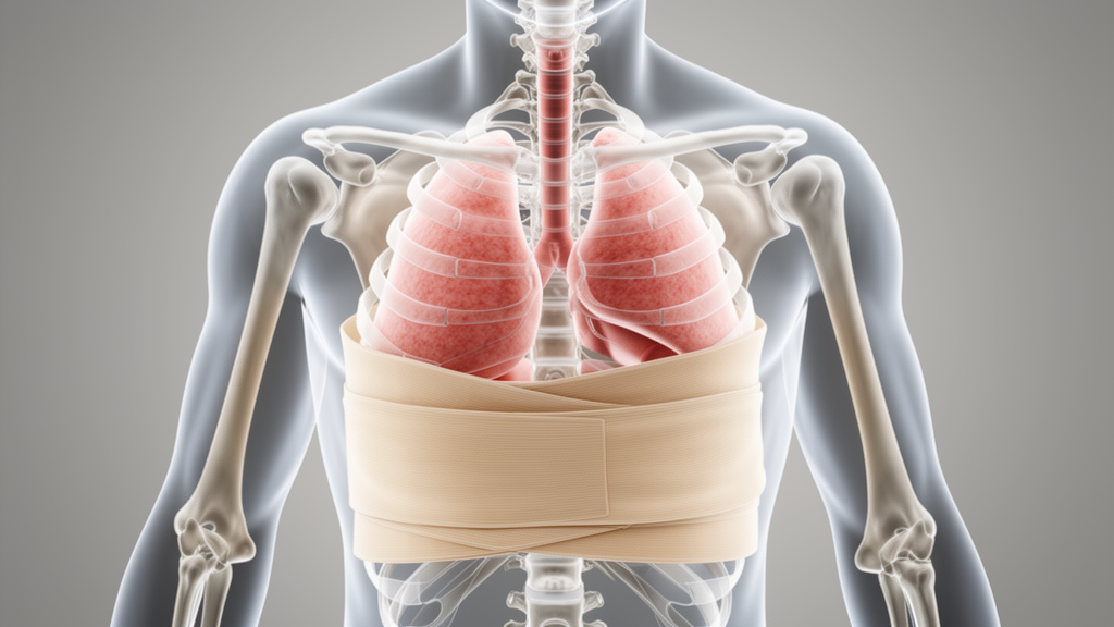
Every breath a person takes depends on the unhindered movement of the chest wall, a living bellows that expands and contracts thousands of times a day to keep the balance of oxygen and carbon dioxide in the blood in check. When that movement is restricted, whether by disease, external equipment, or environmental forces, the consequences ripple through the entire body, from impaired gas exchange to cognitive decline and, in extreme cases, loss of consciousness. Yet studying exactly how and why chest wall restriction disrupts breathing has long been hampered by a lack of suitable tools. Now, a team of engineers and physiologists at the University of Florida has built a device that promises to change that, offering researchers an unprecedented ability to simulate, measure, and control the forces that restrict breathing.
The device, described in a recent paper in Frontiers in Medical Engineering, is the brainchild of Victoria Ribeiro Rodrigues, Lizuannette Mejia, Rafael G. Zucchi, Paul W. Davenport, and Nicholas J. Napoli, working across the university’s Human Informatics and Predictive Performance Optimization (HIPPO) Lab and the Department of Physiological Sciences. Their goal was ambitious: to create a single apparatus capable of applying both static and dynamic loads to the chest wall and abdomen, with precise, reproducible, and independently adjustable force levels. Existing methods, they argue, have fallen short on all these fronts, forcing researchers to choose between crude approximations that lack quantitative feedback or elaborate setups that cannot transition between restriction types.
At the heart of the problem is a fundamental distinction between two kinds of restriction. Static restriction immobilizes the chest wall entirely, regardless of how hard a person tries to breathe, mimicking conditions such as external bracing or severe mechanical constraint. Dynamic restriction, by contrast, applies a constant resistive load that allows movement only when the breathing muscles generate sufficient force to overcome it, a scenario that better reflects the challenges faced by people with chronic obstructive pulmonary disease (COPD), jet pilots enduring high g-forces, or race car drivers under sustained acceleration. Prior devices have typically addressed one or the other, but never both within a single, instrumented platform.
The University of Florida team’s solution is a wooden frame structure, roughly 48 inches tall and 33 inches wide, that surrounds a seated participant. Two middle boards, one positioned over the chest and one over the abdomen, slide along four rods connecting a padded front compression surface to a rear support board. Each middle board carries a vinyl strapping band, three feet long and eight inches wide, that wraps around the participant’s torso and secures with Velcro. A ratchet gear mechanism tightens the bands, pulling them snug against the body, while an adjustable width system of sliding wooden blocks ensures the bands conform to participants of different sizes. The vinyl material is deliberately non-stretchable, preventing any unintended give that could confound measurements.
What elevates the device beyond its predecessors is its instrumentation. Two Linear Variable Differential Transducers, or LVDTs, supplied by Omega, measure the horizontal displacement of the chest wall and abdomen during breathing, capturing even subtle changes in movement patterns. Pressure sensors, built around Millar Mikro-Cath pressure catheters inserted into air bladders filled with exactly 60 milliliters of air, monitor the force exerted by the bands on the chest and abdomen. Before each trial, the catheters are calibrated according to manufacturer guidelines, and a live data feed allows researchers to verify that the applied load matches the target pressure, tightening the bands further if the baseline falls short.
The transition between static and dynamic modes is elegantly simple. In static mode, four shaft collars are positioned and tightened on the rods, locking the middle boards in place and preventing any forward movement regardless of the participant’s effort. In dynamic mode, the collars are removed and constant force springs, flat strips of spring material coiled into cylinders, are mounted on the backboard and connected to the middle boards via carabiners. Unlike conventional compression springs, these provide a steady force throughout their range of motion, reaching full strength after being stretched to 1.25 times their diameter. This allows the participant to move the boards slightly while maintaining a consistent resistive load, simulating the kind of graded restriction encountered in real-world scenarios.
To validate the device, the team recruited six healthy, non-smoking volunteers, four males and two females, with a mean age of 24 years and a mean body mass index of 25.56 kilograms per square meter. Participants underwent pulmonary function testing under three conditions: baseline without restriction, static restriction, and dynamic restriction with a load of 45 kilograms. The tests measured forced expiratory volume in one second (FEV1) and forced vital capacity (FVC), following American Thoracic Society and European Respiratory Society acceptability criteria. Respiratory data, including airflow and mouth pressure, were recorded using a pneumotach connected to a mouthpiece and a two-way non-rebreathing valve.
The results were striking. Under both static and dynamic restriction, FEV1 and FVC dropped significantly compared to baseline, confirming that the device successfully induces physiologically meaningful restriction. FVC declined by 20.8 percent under dynamic loading and 28.2 percent under static loading, while FEV1 fell by 21.6 percent and 28.2 percent respectively. These reductions align closely with average decreases reported in prior chest wall strapping studies, which typically report FVC reductions of around 25.1 percent and FEV1 reductions of 24.9 percent. Additional metrics, including inspiratory volume, inspiratory time, peak inspiratory airflow, and peak inspiratory pressure, also showed significant differences across conditions, with participants adapting their breathing patterns under load by taking shallower, longer breaths that reduce the instantaneous work of breathing.
Displacement data from the LVDTs provided further confirmation of the device’s capabilities. During static trials, chest and abdominal movement was virtually zero, validating the rigid, non-yielding nature of the restriction. In dynamic trials, displacement reached approximately 0.5 centimeters, demonstrating that the device permits measurable movement under load. Pressure variability tests, conducted across three repeated trials for each condition, showed consistent mean pressures: in the static condition, minimum chest pressures ranged from 3.18 to 3.41 kilopascals with standard deviations between 0.17 and 0.22, while minimum abdominal pressures ranged from 2.70 to 2.96 kilopascals. In the dynamic condition, pressures were similarly reproducible, confirming that the device can deliver adjustable and repeatable loads across participants and sessions.
The implications extend well beyond the laboratory. The researchers envision applications in studying respiratory diseases such as asthma and cystic fibrosis, where chest wall mechanics play a critical role in disease progression. The device could also illuminate the physiological challenges faced by military personnel wearing body armor, firefighters in heavy gear, or astronauts and deep-sea divers operating under unusual pressure conditions. Because the apparatus allows independent restriction of the chest and abdomen, it opens new avenues for understanding how each region contributes to breathing patterns and how the body adapts when one or both are compromised. The team also suggests the platform could be extended to investigate cognitive function, muscle fatigue, and blood circulation under restricted breathing, domains where the interplay between respiratory mechanics and broader physiology remains poorly understood.
Looking ahead, the researchers acknowledge that their validation was conducted in a young adult sample, and that the effects of chest wall restriction may be amplified in older individuals, whose chest walls are stiffer due to calcified cartilage and vertebral deformities, and whose lungs have lost elastic recoil. Future studies, they argue, should include broader age ranges and populations with respiratory diseases, with careful attention to load levels that avoid triggering bronchoconstriction in vulnerable participants. Refinements to accommodate extreme body types and adjustable spring resistance for those with compromised pulmonary function could further expand the device’s reach. For now, the device stands as a versatile new tool in the growing effort to understand, model, and ultimately mitigate the respiratory challenges that arise when the simple act of breathing becomes a battle against constraint.
Subject of Research: Respiratory mechanics and biomedical engineering
Article Title: Chest wall restriction device for modeling respiratory challenges and dysfunction
Article References: Ribeiro Rodrigues, V., Mejia, L., Zucchi, R. G., Davenport, P. W., & Napoli, N. J. (2025). Chest wall restriction device for modeling respiratory challenges and dysfunction. Frontiers in Medical Engineering, 3, Article 1560136. https://doi.org/10.3389/fmede.2025.1560136
Image Credits: AI Generated
DOI: 10.3389/fmede.2025.1560136
Keywords: chest wall restriction, respiratory mechanics, breathing dysfunction, pulmonary function testing, biomedical engineering, static and dynamic loads, COPD, extreme environments, Chest, wall, restriction, device
Cite Scienmag News
Denise Maddox. (October 17, 2026). Chest wall restriction device for modeling respiratory challenges and dysfunction. Scienmag. https://scienmag.com/chest-wall-restriction-device-for-modeling-respiratory-challenges-and-dysfunction/
Denise Maddox. "Chest wall restriction device for modeling respiratory challenges and dysfunction." Scienmag, 17 October 2026, https://scienmag.com/chest-wall-restriction-device-for-modeling-respiratory-challenges-and-dysfunction/. Accessed 17 October 2026.
Denise Maddox. "Chest wall restriction device for modeling respiratory challenges and dysfunction." Scienmag. October 17, 2026. https://scienmag.com/chest-wall-restriction-device-for-modeling-respiratory-challenges-and-dysfunction/


