


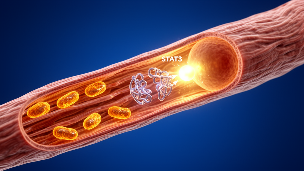
Deep inside heart muscle cells, a molecular editor called OTUD5 is quietly rewriting the chemical tags that govern how the heart responds to chronic stress, and a new study suggests that boosting this editor could help protect the heart from a damaging condition that affects millions of people worldwide. In research published in Experimental & Molecular Medicine, a team led by Bozhi Ye and Yucheng Jiang of Wenzhou Medical University reports that OTUD5, an enzyme belonging to the deubiquitinase family, acts as a powerful brake on pathological cardiac hypertrophy, the abnormal enlargement of heart muscle cells that gradually drives the heart toward failure. The work reveals an unexpected regulatory axis connecting protein editing, transcription factor activity, and mitochondrial energy metabolism, and it identifies a potential new target for treating heart disease in aging populations.
Pathological cardiac hypertrophy is a growing public health challenge, particularly as societies age. When the heart is subjected to chronic stress, such as sustained high blood pressure or hormonal overload, individual myocardial cells increase in size rather than proliferating in number. This compensatory response initially helps the heart maintain contractility, but over time it becomes maladaptive, leading to structural remodeling, fibrosis, and eventually heart failure. The condition is now recognized as an independent risk factor for elevated cardiovascular disease incidence and mortality, underscoring the urgent need for novel therapeutic targets that can intervene at the molecular level before irreversible damage occurs.
To understand how OTUD5 fits into this picture, it helps to grasp the biology of ubiquitination, the process OTUD5 reverses. Ubiquitin is a small protein that cells attach to other proteins as a chemical tag, and these tags come in different chain types that convey different instructions. K48-linked ubiquitin chains typically mark proteins for destruction by the proteasome, whereas K63-linked chains preferentially regulate protein activity and function without affecting stability. Deubiquitinases are enzymes that recognize and cleave these chains, thereby precisely tuning the stability and function of their substrate proteins. OTUD5 is known to cleave both K48- and K63-linked chains, and previous studies have implicated it in DNA damage repair, immune regulation, inflammatory responses, and cell death, including a protective role against ferroptosis during myocardial ischemia-reperfusion injury.
The research team first examined OTUD5 expression in mouse models of pathological cardiac hypertrophy, using both angiotensin II infusion and transverse aortic constriction, a surgical procedure that creates chronic pressure overload. They found that OTUD5 mRNA and protein levels were significantly upregulated in the hearts of mice subjected to either treatment compared with controls. Human heart samples from patients diagnosed with heart failure confirmed that OTUD5 mRNA was elevated in hypertrophic myocardial tissues. Immunofluorescence staining revealed that the increased OTUD5 was predominantly localized within cardiomyocytes rather than fibroblasts or macrophages, suggesting a cardiomyocyte-specific expression pattern. In vitro experiments with neonatal rat cardiomyocytes showed that OTUD5 protein increased in a time-dependent manner following angiotensin II stimulation, while angiotensin II failed to induce OTUD5 expression in primary cardiac fibroblasts or macrophages.
To dissect the functional role of this cardiomyocyte-enriched enzyme, the researchers generated a cardiomyocyte-specific OTUD5 knockout mouse model by crossing floxed OTUD5 mice with Myh6-Cre transgenic mice. When these knockout mice were infused with angiotensin II for four weeks, left ventricular hypertrophy became more pronounced than in littermate controls, as evidenced by increased anterior wall and ventricular septal thickness at end-diastole, reduced ejection fraction, and reduced fractional shortening on echocardiography. Heart weight-to-body weight and heart weight-to-tibial length ratios increased significantly, histological analyses showed enlarged cardiomyocyte cross-sectional areas, and Masson’s trichrome and Sirius Red staining revealed more severe myocardial fibrosis. Molecular markers of hypertrophy, including myosin heavy chain and atrial natriuretic peptide, were further elevated in the knockout hearts. A similar pattern emerged under transverse aortic constriction, with OTUD5 deficiency exacerbating cardiac dysfunction, hypertrophy, fibrosis, and hypertrophic gene expression.
Conversely, when the researchers delivered OTUD5 overexpression specifically to cardiomyocytes using an adeno-associated virus serotype 9 vector carrying the cardiac troponin T promoter, they observed significant protection against angiotensin II-induced cardiac dysfunction. Ejection fraction and fractional shortening increased compared with the control group, and the therapeutic effects emerged in a time-dependent manner, becoming statistically significant at week four. Histological analyses confirmed that OTUD5 overexpression effectively mitigated angiotensin II-induced myocardial hypertrophy and fibrosis, along with reduced serum atrial natriuretic peptide levels. These results establish that cardiomyocyte OTUD5 acts as a protective factor during pathological cardiac remodeling.
To identify the molecular substrate through which OTUD5 exerts this protection, the team performed co-immunoprecipitation combined with liquid chromatography-tandem mass spectrometry on HL-1 cells overexpressing Flag-tagged OTUD5 under angiotensin II stimulation. Among the interacting proteins identified, STAT3 stood out because it is known to participate directly in cardiac hypertrophy. Subsequent co-immunoprecipitation assays confirmed an endogenous interaction between OTUD5 and STAT3 in angiotensin II-stimulated cardiac tissue, while OTUD5 did not directly bind to other candidate substrates such as SPTBN1 or SORBS3. Further experiments demonstrated that OTUD5 overexpression considerably reduced STAT3 ubiquitination, specifically through a K63-linked manner, without affecting K48-linked ubiquitination or total STAT3 protein levels, indicating that OTUD5 modulates STAT3 activity rather than stability.
Sequence analysis revealed that the catalytic active site of OTUD5 is located at a highly conserved cysteine residue, C224. When the researchers mutated this residue to serine, the mutant completely lost its capacity to remove ubiquitin chains from STAT3. Using a ubiquitination peptide enrichment assay, they identified lysine 283 and lysine 707 as potential deubiquitination sites on STAT3. Functional assays showed that the K283R mutation significantly attenuated angiotensin II-induced hypertrophic gene expression in HL-1 cells, whereas the K707R mutation did not. The K283R variant exhibited markedly reduced ubiquitination levels compared with wild-type STAT3, and this modification was resistant to further reduction by OTUD5, establishing K283 as the primary site for OTUD5-mediated STAT3 deubiquitination. Notably, K283 resides within the DNA-binding domain of STAT3, spatially distant from the SH2 domain responsible for receiving JAK2 signals, suggesting that K283 ubiquitination likely modulates the JAK2-STAT3 interaction through an allosteric mechanism rather than direct physical interference.
STAT3 activation requires phosphorylation at tyrosine 705 and subsequent nuclear translocation. The researchers found that silencing OTUD5 in HL-1 cells markedly exacerbated angiotensin II-induced STAT3 phosphorylation, whereas OTUD5 overexpression considerably attenuated it. Nuclear fraction analysis confirmed that OTUD5 silencing increased STAT3 nuclear accumulation, while overexpression suppressed this process. Co-immunoprecipitation assays demonstrated that OTUD5 overexpression markedly weakened the interaction between JAK2, the upstream kinase, and STAT3, and crucially, the K283R mutation completely abrogated this regulatory effect. Loss-of-function assays revealed that silencing STAT3 not only mitigated angiotensin II-induced hypertrophy but also negated the protective phenotype conferred by OTUD5 overexpression, confirming that STAT3 mediates the protective effects of OTUD5 in cardiomyocytes.
As a transcription factor, STAT3 regulates gene expression programs downstream of its activation. RNA sequencing of transverse aortic constriction-challenged mouse hearts with or without OTUD5 knockout revealed a notable downregulation of PGC1α, the master regulator of mitochondrial biogenesis and energy metabolism, in the knockout hearts. PGC1α orchestrates the transcriptional activation of genes involved in oxidative phosphorylation and mitochondrial function by coactivating nuclear receptors such as peroxisome proliferator-activated receptors, estrogen-related receptors, and nuclear respiratory factors. The researchers confirmed that OTUD5 positively regulates Pgc1α gene transcription through STAT3 using CUT&Tag assays, which measure STAT3 DNA-binding activity at the Pgc1α locus. Silencing OTUD5 reduced PGC1α expression, while overexpression upregulated it, and PGC1α protein levels were significantly diminished in myocardial tissues of OTUD5 knockout mice challenged with either angiotensin II or transverse aortic constriction.
Mitochondrial respiratory function was evaluated using a Seahorse XFe96 Analyzer to measure oxygen consumption rates in HL-1 cells. The results indicated that OTUD5 overexpression significantly mitigated the angiotensin II-induced elevation in mitochondrial oxidative phosphorylation, as evidenced by enhanced basal respiration, ATP production-coupled respiration, and maximal respiratory capacity. Importantly, silencing STAT3 completely abolished these protective effects of OTUD5. JC-1 staining confirmed that angiotensin II exposure led to significant mitochondrial depolarization in OTUD5-silenced cells, whereas OTUD5 overexpression markedly attenuated this phenotype. These findings reveal that OTUD5 prevents mitochondrial dysfunction by inhibiting STAT3 activity and thereby upregulating PGC1α expression in cardiomyocytes.
To validate the role of STAT3 as a functional substrate of OTUD5 in vivo, the researchers administered OTUD5 overexpression to cardiomyocyte-specific knockout mice via tail vein injection, followed by oral delivery of Stattic, a STAT3-specific inhibitor. These mice were then infused with angiotensin II for four weeks. Echocardiographic analysis demonstrated that either OTUD5 restoration or STAT3 inhibition significantly improved cardiac function in angiotensin II-infused knockout mice. However, OTUD5 overexpression following STAT3 inhibition did not result in further improvement, confirming that STAT3 is the critical mediator of OTUD5’s protective effects. Histological assessments, including measurements of cardiac size, hematoxylin and eosin staining, wheat germ agglutinin staining, Masson’s trichrome staining, and Sirius Red staining, revealed that Stattic effectively mitigated myocardial hypertrophy and fibrosis, and OTUD5 overexpression provided no additional benefit in Stattic-treated mice.
The discovery that OTUD5 uniquely targets STAT3 at lysine 283 within its DNA-binding domain distinguishes this enzyme from other deubiquitinases that regulate STAT3 through degradation-associated sites. Unlike USP28, OTUB1, and YOD1, which remove K48-linked ubiquitination affecting STAT3 stability, or CYLD, TRAF6, OTUD1, and USP20, which deubiquitinate STAT3 in a K63-linked manner to influence phosphorylation or nuclear translocation, OTUD5 selectively removes K63-linked chains to modulate STAT3 transcriptional activity without compromising protein stability. Functionally, OTUD5 alleviates STAT3-mediated repression of PGC1α and enhances mitochondrial bioenergetics, establishing a regulatory axis not previously reported for other deubiquitinating enzymes. This finding provides fresh insights into developing precise modulators for STAT3 activity, a target that has proven challenging because STAT3 lacks a conventional active site and shares high homology with other STAT family members.
Currently, no small molecule agonists or activators targeting OTUD5 have been reported, and strategies for enhancing OTUD5 function rely entirely on adeno-associated virus-mediated gene overexpression. This approach has demonstrated notable preclinical efficacy in various disease models, including diabetic nephropathy, myocardial ischemia-reperfusion injury, and acute kidney injury. The researchers suggest that the C224 catalytic site and the adjacent substrate-binding groove in the OTUD5 structure could be used in drug design to construct allosteric small-molecule agonists or molecular adhesives that enhance endogenous OTUD5 activity and improve pathological myocardial remodeling. Future research directions include clarifying whether OTUD5 regulates other mitochondrial-associated transcription factors, delineating the broader downstream cascades of STAT3 using chromatin immunoprecipitation sequencing and transcriptomic analyses, and advancing cardiomyocyte-specific delivery methods for OTUD5 while validating the safety profiles of OTUD5-overexpressing therapies. The study establishes a comprehensive molecular cascade connecting deubiquitination, STAT3 transcriptional regulation, PGC1α expression, and mitochondrial bioenergetics, offering potential targets for therapeutic interventions combating heart failure through the OTUD5-STAT3-PGC1α axis.
Subject of Research: Cardiomyocyte-enriched OTUD5 alleviates pathological myocardial hypertrophy by deubiquitinating STAT3 and modulating the STAT3–PGC-1α axis
Article Title: Cardiomyocyte-enriched OTUD5 alleviates pathological myocardial hypertrophy by deubiquitinating STAT3 and modulating the STAT3–PGC-1α axis
Article References: Ye, B., Jiang, Y., Zheng, Z., Jin, Y., Liao, Y., Jia, Z., Lin, W., Xu, D., Han, J., Fang, Z., Zhang, N., Shan, P., Wu, G., & Liang, G. (2026). Cardiomyocyte-enriched OTUD5 alleviates pathological myocardial hypertrophy by deubiquitinating STAT3 and modulating the STAT3–PGC-1α axis. Experimental & Molecular Medicine. https://doi.org/10.1038/s12276-026-01870-5
Image Credits: AI Generated
DOI: 10.1038/s12276-026-01870-5
Keywords: Cardiomyocyte-enriched, OTUD5, alleviates, pathological, myocardial, hypertrophy, deubiquitinating, STAT3, modulating, PGC-1, axis, scientific research
Cite Scienmag News
Nathaniel Bowman. (October 16, 2026). Cardiomyocyte-enriched OTUD5 alleviates pathological myocardial hypertrophy by deubiquitinating STAT3 and modulating the STAT3–PGC-1α axis. Scienmag. https://scienmag.com/cardiomyocyte-enriched-otud5-alleviates-pathological-myocardial-hypertrophy-by-deubiquitinating-stat3-and-modulating-the-stat3-pgc-1%ce%b1-axis/
Nathaniel Bowman. "Cardiomyocyte-enriched OTUD5 alleviates pathological myocardial hypertrophy by deubiquitinating STAT3 and modulating the STAT3–PGC-1α axis." Scienmag, 16 October 2026, https://scienmag.com/cardiomyocyte-enriched-otud5-alleviates-pathological-myocardial-hypertrophy-by-deubiquitinating-stat3-and-modulating-the-stat3-pgc-1%ce%b1-axis/. Accessed 16 October 2026.
Nathaniel Bowman. "Cardiomyocyte-enriched OTUD5 alleviates pathological myocardial hypertrophy by deubiquitinating STAT3 and modulating the STAT3–PGC-1α axis." Scienmag. October 16, 2026. https://scienmag.com/cardiomyocyte-enriched-otud5-alleviates-pathological-myocardial-hypertrophy-by-deubiquitinating-stat3-and-modulating-the-stat3-pgc-1%ce%b1-axis/


