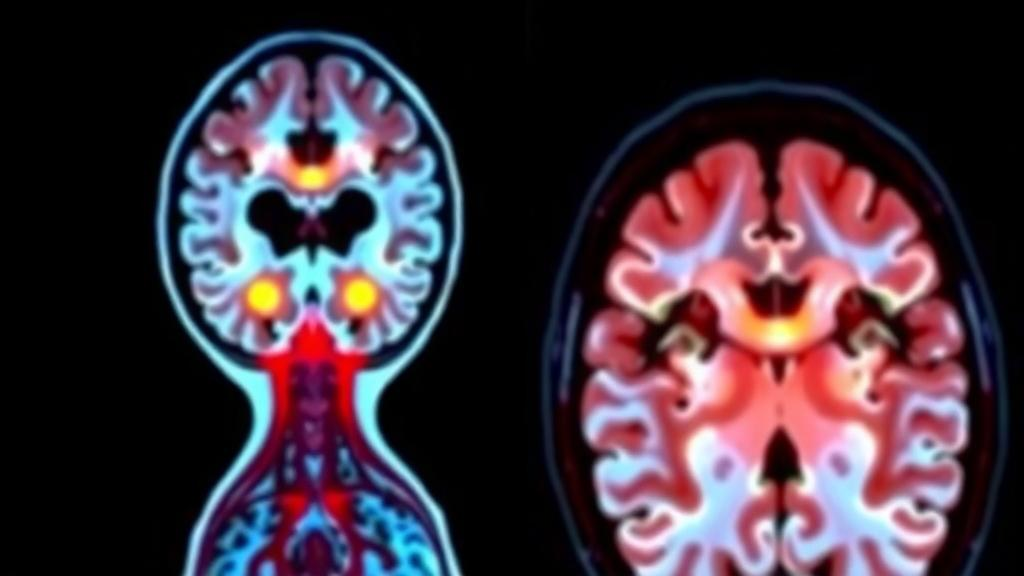
In a finding that could reshape how clinicians assess one of the most stubborn complications of Crohn’s disease, researchers in China have shown that patterns of brain activity and structure, captured with a standard MRI scanner, can help gauge how severely a patient’s intestine is stiffening with fibrotic scar tissue. The study, conducted at two centers and published in BMC Medical Imaging, suggests that the long-theorized gut-brain axis is not merely a conduit for inflammation and pain signaling but may leave measurable fingerprints in the brain that mirror what is happening deep within the bowel wall.
Intestinal fibrosis is among the most feared consequences of Crohn’s disease. Over years of chronic inflammation, the bowel wall accumulates excess extracellular matrix, thickens, and loses its elasticity, eventually producing strictures that can obstruct the passage of food and often require surgical resection. Unlike inflammation, fibrosis does not reliably respond to the biologic therapies that have transformed Crohn’s care, and detecting it early remains a major clinical challenge. Current assessment relies on magnetic resonance enterography, in which the small bowel is distended and imaged, sometimes supplemented by magnetization transfer imaging, a technique sensitive to macromolecular content in tissue that can hint at fibrotic change. But these methods interrogate the bowel itself, and their findings can be limited by distension quality, motion, and interpretation variability.
The new research took an unconventional approach: instead of looking harder at the gut, the team looked at the brain. Led by Xiaodi Shen, Qingzhu Zheng, and Dailin Li, with corresponding authors Ruonan Zhang, Qiaochu Zhao, and Lili Huang at The First Affiliated Hospital of Sun Yat-Sen University in Guangzhou, the prospective two-center study enrolled 109 participants. Each underwent a battery of multiparametric brain MRI covering functional, structural, and microstructural dimensions of neural tissue, alongside magnetic resonance enterography with magnetization transfer imaging and a targeted serum metabolomics panel measuring 429 metabolites. The goal was ambitious: to build a machine-learning model, a so-called brain fingerprint, that could distinguish patients with moderate-to-severe intestinal fibrosis from those with non-to-mild fibrosis using brain imaging alone.
The technical machinery behind the fingerprint is worth unpacking. The researchers drew on several complementary MRI contrasts. Functional measures included the amplitude of low-frequency fluctuations, or ALFF, which quantifies the intensity of spontaneous blood-oxygenation-level-dependent signal oscillations at rest, and regional homogeneity, which captures the local synchrony of neural activity. Microstructural measures probed the integrity of neural tissue at the cellular level, including cortical R2*, a relaxation metric sensitive to tissue iron and myelination, and diffusion-based parameters that reflect neurite density and orientation dispersion. Structural and functional features from distributed cortical and subcortical regions were then fed into a recursive feature elimination pipeline, an embedded machine-learning feature selection strategy that iteratively discards the least informative variables, ultimately distilling the model to 13 neuroimaging features.
The performance of the resulting brain fingerprint index was striking. In the training set it achieved an area under the receiver operating characteristic curve of 0.923, and in independent validation and test cohorts it held up at 0.837 and 0.846 respectively. An AUC in the mid-0.80s on unseen data indicates discrimination well above chance and approaching the range where a biomarker could meaningfully complement existing tools, particularly one derived entirely from extraintestinal imaging. The model was trained to separate patients classified as BF2, meaning moderate-to-severe bowel fibrosis, from those classified as BF1, meaning non-to-mild fibrosis, with fibrosis severity defined by normalized magnetization transfer ratio findings on enterography.
Beyond classification, the brain fingerprint index showed meaningful correlations with bowel-level measures. It correlated positively with normalized magnetization transfer ratio, with a correlation coefficient of 0.53, suggesting that as the brain signature intensified, the bowel tissue showed greater macromolecular change consistent with fibrogenesis. The index also correlated with the presence of bowel stricture, with a coefficient of 0.31, a more modest but still notable association given the biological distance between the two organ systems. These correlations imply that the neural signature is not an arbitrary statistical artifact but tracks, at least in part, the severity of structural change in the intestine.
What exactly differs in the brains of patients with more severe fibrosis? The study found that patients with moderate-to-severe fibrosis exhibited reduced cortical R2* values and reduced amplitude of low-frequency fluctuations, alongside increased activity in the isthmus of the cingulate cortex. The cingulate cortex is a hub of the brain’s pain and interoceptive networks, regions that integrate signals from the body’s interior and are known to be altered in chronic visceral pain conditions. Reduced spontaneous activity across broader cortex, paired with heightened engagement of this interoceptive hub, paints a picture of a nervous system reorganized by chronic gut disease, though the direction of causality remains an open question. The brain changes could reflect the cumulative burden of visceral pain and inflammation, or they could participate in the feedback loops that modulate gut immune and stromal responses.
To probe the biochemical bridge between brain and bowel, the team performed exploratory mediation analyses linking brain imaging features, serum metabolites, and fibrosis-associated enterography findings. These analyses yielded 13 nominal indirect associations involving circulating metabolites, hinting at possible metabolic mediators of the gut-brain relationship. However, none of these associations survived correction for multiple comparisons using the Benjamini-Hochberg false discovery rate procedure, a statistical safeguard against spurious findings when many hypotheses are tested simultaneously. The authors are appropriately cautious on this point: the metabolite-mediated pathways remain hypotheses for future work rather than established mechanisms. Still, the framework itself, integrating neuroimaging, enterography, and metabolomics in the same patients, offers a template for how multi-omics and multi-organ imaging can be combined to dissect complex systemic diseases.
The clinical implications, if the findings are replicated, could be substantial. A brain MRI-based marker would be noninvasive in the fullest sense, requiring no bowel preparation, no enteric tube, and no distension of the gut. It could potentially be obtained during routine brain imaging or incorporated into existing MRI sessions, offering an extraintestinal window on fibrosis severity that complements, rather than replaces, direct bowel imaging. For patients in whom enterography is technically difficult, poorly tolerated, or contraindicated, a complementary neural marker could help triage who needs more aggressive bowel-directed workup. The approach might also prove useful in monitoring disease progression over time, since fibrosis evolves slowly and repeated enterographies carry cumulative burden for patients.
Important caveats temper the excitement. The study is cross-sectional, capturing a snapshot rather than tracking individuals over time, so it cannot establish whether brain changes precede, accompany, or follow fibrotic progression. The sample of 109 participants, while respectable for a multimodal imaging study, will need expansion in larger and more diverse cohorts, and external validation at additional centers will be essential before any clinical deployment. The mediation findings did not survive statistical correction, leaving the mechanistic story incomplete. And the fibrosis reference standard itself, magnetization transfer ratio-defined severity on enterography, is an imaging surrogate rather than histologic gold standard. Nevertheless, the study represents a genuinely novel demonstration that multiparametric brain MRI features are associated with intestinal fibrosis severity in Crohn’s disease, and that a compact 13-feature neural fingerprint can stratify patients with robust discrimination. As the authors suggest, the brain fingerprint may provide a noninvasive extraintestinal imaging marker complementary to local bowel imaging, a concept that, if validated, would mark a striking convergence of neurology and gastroenterology in the service of patients with chronic inflammatory bowel disease.
Subject of Research: Using multiparametric brain MRI and machine learning to stratify intestinal fibrosis severity in Crohn's disease via the gut-brain axis
Article Title: Multiparametric brain MRI fingerprints stratify intestinal fibrosis severity in Crohn’s disease
Article References: Shen, X., Zheng, Q., Li, D., Zheng, W., Zhuang, X.-Z., Fan, C., Wang, Y., Li, X., Zhang, R., Zhao, Q., & Huang, L. (2026). Multiparametric brain MRI fingerprints stratify intestinal fibrosis severity in Crohn’s disease. BMC Medical Imaging. https://doi.org/10.1186/s12880-026-02795-1
Image Credits: AI Generated
DOI: 10.1186/s12880-026-02795-1
Keywords: Crohn's disease, intestinal fibrosis, brain MRI, gut-brain axis, machine learning, magnetic resonance enterography, magnetization transfer ratio, brain fingerprint, ALFF, serum metabolomics, bowel stricture, BMC Medical Imaging
Cite Scienmag News
Cassandra Pierce. (October 11, 2026). Brain Scans Reveal Hidden Clues to Gut Scarring in Crohn’s Disease. Scienmag. https://scienmag.com/brain-scans-reveal-hidden-clues-to-gut-scarring-in-crohns-disease/
Cassandra Pierce. "Brain Scans Reveal Hidden Clues to Gut Scarring in Crohn’s Disease." Scienmag, 11 October 2026, https://scienmag.com/brain-scans-reveal-hidden-clues-to-gut-scarring-in-crohns-disease/. Accessed 11 October 2026.
Cassandra Pierce. "Brain Scans Reveal Hidden Clues to Gut Scarring in Crohn’s Disease." Scienmag. October 11, 2026. https://scienmag.com/brain-scans-reveal-hidden-clues-to-gut-scarring-in-crohns-disease/