


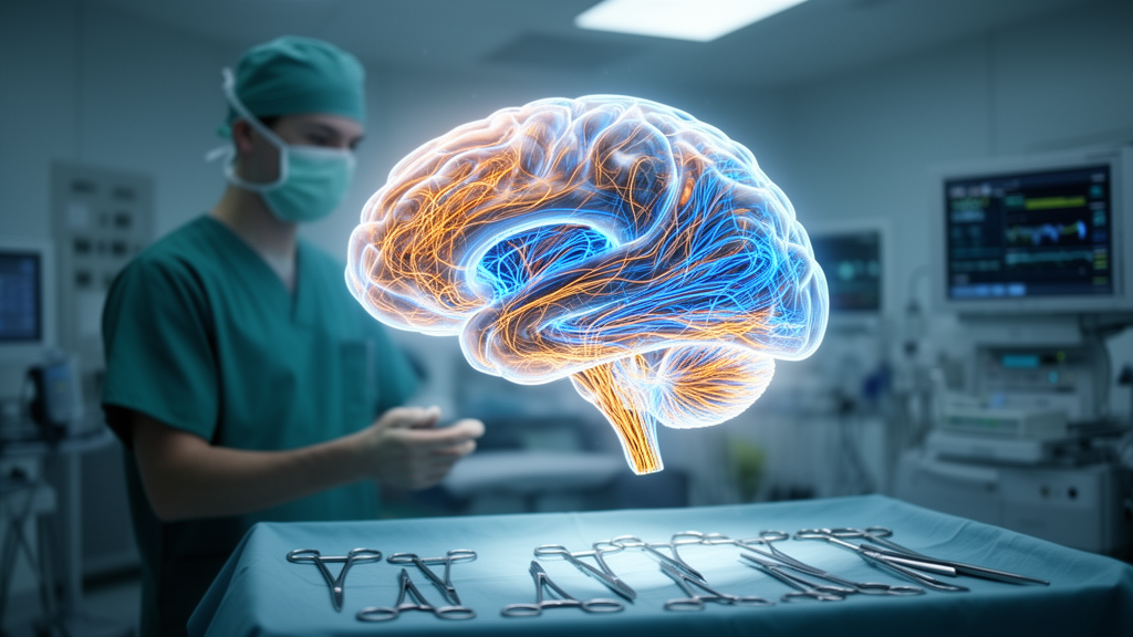
For patients with gliomas in the temporal lobe, the operation itself can sometimes feel like a second diagnosis. Surgeons may successfully remove the tumour, yet patients walk out of the hospital with memory lapses, attention deficits, and language difficulties that reshape their daily lives. A new study published in BMC Medical Imaging suggests that the problem may lie not only in the tumour or in the classic functional areas that surgeons already try to spare, but in the large-scale brain networks that connect those areas. By mapping each patient’s functional connectome before surgery, a team of Chinese neurosurgeons and neuroscientists reports that they could see tumour invasion spreading through cognitive networks, redesign the surgical plan accordingly, and measurably improve both the protection of brain structures and patients’ cognitive scores after the operation.
The research, led by Zhiqiang Wu, Jiacheng Lu, Hao You, and Siyan Hou with corresponding authors Junxia Zhang and Qin Ye, draws on connectomics, the field devoted to mapping the structural and functional topology of the human brain as a unified network. Rather than viewing the brain as a collection of isolated regions with fixed jobs, connectomics treats it as a set of interacting networks. The study focused on five of the most important of these: the salience network, which helps the brain decide which stimuli deserve attention; the central executive network, which supports planning and decision-making; the default mode network, active during introspection and memory retrieval; and the dorsal and ventral attention networks, which orient attention to the external world. All five have critical hubs in or near the temporal lobes, which is precisely where temporal gliomas grow.
To build these personalized network maps, the team used imaging data from each patient, applying techniques associated with the Human Connectome Project and diffusion tractography imaging, which traces the orientation of water molecules in brain tissue to infer the paths of white matter fibres. The approach allowed the researchers to reconstruct not only grey-matter functional structures but also the major fibre bundles that carry signals between them, including the inferior fronto-occipital fasciculus, the inferior longitudinal fasciculus, and the optic radiations. These tracts are the wiring of the brain’s long-distance communication, and damaging them can sever the connections that cognitive networks depend on even when the cortical areas themselves appear intact.
In the retrospective portion of the study, the researchers analysed 53 patients with temporal gliomas whose personalized functional networks had been reconstructed from imaging. The findings were striking. In 52 of the 53 patients, or 98.11 percent, tumour infiltration extended into brain networks and fibre tracts, a slightly higher rate than the 94.34 percent of patients whose tumours invaded discrete functional structures. In other words, nearly every tumour was not simply a local mass pressing on a language area or a memory hub; it was a lesion woven into the connective fabric of the brain. This network-level invasion, largely invisible to conventional surgical planning that focuses on landmarks and eloquent cortex, offers a plausible explanation for why cognitive decline persists after technically successful resections.
The second phase of the study moved from observation to intervention. The researchers compared 29 patients with temporal gliomas operated on with connectomics-optimized surgical programs, the experimental group, against patients treated with traditional surgical strategies, the control group. Before surgery, the two groups showed no significant differences in the characteristics of tumour invasion, meaning the comparison was not skewed by one group having more aggressive or more widespread disease. After the operations, the differences became clear. In the 16 postoperative patients in the experimental group, the protection rates for brain networks and for functional structures were noticeably higher than in the 13 patients who underwent traditional surgeries, with statistical significance reported at P = 0.000 for network protection and P = 0.029 for functional structure protection.
The left temporal lobe deserves special mention because it houses much of the language machinery of the brain in most people, making surgery there particularly delicate. When the analysis was restricted to the 18 patients with left temporal gliomas, the pattern held. The eight patients in the connectomics-guided experimental group had significantly greater protection of both brain networks and functional structures than the ten patients in the control group, with P values of 0.017 and 0.029 respectively. Both surgical outcomes and cognitive scores, assessed with tools such as the Montreal Cognitive Assessment, were significantly better in the connectomics group than in the traditional treatment group, with P values below 0.05 across these comparisons.
Technically, the connectomics-guided approach changes what the surgeon sees and plans around. Traditional neurosurgical planning emphasizes avoiding eloquent cortex, the regions where electrical stimulation or functional MRI shows critical functions such as movement, speech, or vision. The connectomic approach adds a second layer: it visualizes the network edges, the white matter pathways and their connections to distributed cognitive systems, and asks the surgeon to preserve the topology of those systems while still achieving maximal tumour resection. In practice, this means an incision trajectory and resection boundary designed not only around what a region does but around what it connects to. A tumour that infiltrates the inferior fronto-occipital fasciculus, for example, threatens the link between occipital visual processing and frontal language and executive areas, and cutting along the tumour margin without accounting for that tract could disconnect a network whose nodes are all perfectly preserved.
The clinical implication, the authors argue, is that preserving traditional functional structures is necessary but no longer sufficient. During temporal glioma surgery, they conclude, it is essential to also retain the brain networks that carry advanced cognitive functions. Their preliminary evidence indicates that connectomics-based visualization of functional networks improves neurosurgical comprehension, optimizes surgical programs, and holds the potential to minimize surgical trauma while enabling superior levels of tumour resection with preserved functional outcomes. For a disease where the balance between oncological control and quality of life is the central clinical tension, that framing reframes the goal of surgery: the target is no longer just the tumour volume but the integrity of the patient’s cognitive connectome.
Several caveats temper the enthusiasm. The comparative analysis involved relatively small numbers of patients, 29 in the overall comparison and 18 in the left temporal subgroup, and the study design was retrospective in its first phase, which limits the strength of causal conclusions. The published version was also shared early as a citable accepted manuscript subject to further edits. Larger, prospective, and ideally randomized comparisons will be needed before connectomics-guided planning becomes a standard of care. The study was conducted under the Declaration of Helsinki with ethics approval from the First Affiliated Hospital with Nanjing Medical University and informed consent from all participants, and the authors declared no competing interests, with funding from a Clinical Capacity Enhancement Project grant at Jiangsu Province Hospital.
Even with those limitations, the study lands at a moment when the tools of network neuroscience are maturing rapidly. Machine learning methods, which the authors list among their keywords, are increasingly able to segment networks, predict cognitive deficits from connectivity patterns, and integrate multimodal imaging into surgical navigation. The idea that a glioma should be understood as an attack on a network rather than an occupation of a region is philosophically simple but surgically transformative, and this work offers an early, quantified demonstration that acting on that idea changes outcomes. If larger trials confirm these results, the connectome may become as routine a preoperative map for neurosurgeons as the anatomical MRI is today, and the devastating trade-off between removing a tumour and losing one’s cognitive self may become considerably less stark.
Subject of Research: Connectomics-guided surgical planning to preserve cognitive brain networks in temporal glioma patients
Article Title: Connectomics-based functional networks in temporal gliomas
Article References: Wu, Z., Lu, J., You, H., Hou, S., Zhang, Z., Ye, M., Wang, X., Yuan, C., Zhang, J., & Ye, Q. (2026). Connectomics-based functional networks in temporal gliomas. BMC Medical Imaging. https://doi.org/10.1186/s12880-026-02715-3
Image Credits: AI Generated
DOI: 10.1186/s12880-026-02715-3
Keywords: connectomics, temporal glioma, brain networks, neurosurgery, cognitive function, default mode network, salience network, central executive network, diffusion tractography, white matter tracts, BMC Medical Imaging, brain mapping
Cite Scienmag News
Ophelia Keating. (October 18, 2026). Brain Network Maps Help Surgeons Protect Cognition in Temporal Glioma Patients. Scienmag. https://scienmag.com/brain-network-maps-help-surgeons-protect-cognition-in-temporal-glioma-patients/
Ophelia Keating. "Brain Network Maps Help Surgeons Protect Cognition in Temporal Glioma Patients." Scienmag, 18 October 2026, https://scienmag.com/brain-network-maps-help-surgeons-protect-cognition-in-temporal-glioma-patients/. Accessed 18 October 2026.
Ophelia Keating. "Brain Network Maps Help Surgeons Protect Cognition in Temporal Glioma Patients." Scienmag. October 18, 2026. https://scienmag.com/brain-network-maps-help-surgeons-protect-cognition-in-temporal-glioma-patients/


