


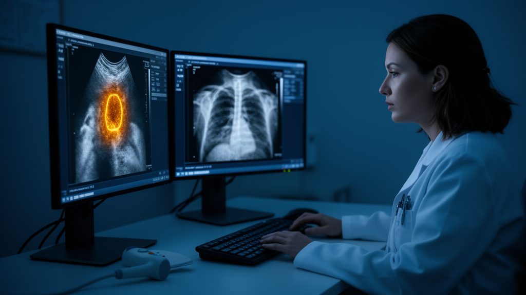
Lung cancer remains one of the deadliest malignancies in the world, and one of its most diagnostically frustrating presentations is the subpleural pulmonary lesion, a growth that sits just beneath the pleural surface of the lung. Because these lesions abut the chest wall, they are actually visible on ultrasound, a cheap, radiation-free, and widely available imaging modality. Yet reading those grayscale images has always been a deeply subjective exercise, dependent on the training and instincts of whoever holds the probe. A new study published in BMC Medical Imaging suggests that subjectivity may soon be a problem of the past. Researchers in Shanghai, working with a collaborator at Emory University, have built a two-stage deep learning framework that can automatically outline subpleural lesions on ultrasound and then judge, with impressive reliability, whether the finding is likely benign or malignant.
The study, led by Qiang Ma and TianXi Liang with corresponding author Yin Wang of Shanghai Pulmonary Hospital, was designed as a retrospective, multicenter analysis. The team assembled ultrasound data from 1,059 patients with subpleural pulmonary lesions, drawn from a primary center between January 2023 and December 2024, with additional external cohorts recruited from two other hospitals between June and December 2024. The patient population had a mean age of roughly 61 years and was predominantly male, with 726 male patients among the 1,059 enrolled. Crucially, the researchers did not simply test their models on data from the same hospital that produced the training set. They held out two entirely separate external test sets, comprising 108 and 94 patients respectively, to see whether the artificial intelligence could generalize to machines, protocols, and patient populations it had never encountered.
The architecture of the framework reflects a logical division of labor. The first stage is segmentation: the computer must find the lesion in the ultrasound image and trace its boundaries precisely. To do this, the team benchmarked three well-established convolutional neural network approaches: nnU-Net, U-Net, and DeepLabv3+. The nnU-Net, a self-configuring variant of the U-Net family that has become something of a gold standard in medical image segmentation, emerged as the clear winner. Its performance was measured with the Dice similarity coefficient, a metric that scores the overlap between the machine-drawn region of interest and the ground-truth annotation, where a value of 1.0 indicates perfect agreement. The nnU-Net achieved a Dice score of 0.920 on the internal tuning-validation set of 257 cases, and, more importantly, held its ground externally, scoring 0.913 on External Test Set 1 and 0.915 on External Test Set 2. Those numbers mean the model’s outlines agreed with expert annotations to within a few percent of perfect overlap, even on unfamiliar data.
With the lesion reliably isolated, the second stage took over: classification. Here the researchers cast a wide net, training and comparing thirteen different candidate architectures drawn from the most influential families of convolutional networks in modern computer vision. The lineup included DenseNet121 and DenseNet201, the EfficientNet family from B0 through B5, three depths of ResNet (18, 50, and 101), Inception-V3, and VGG19. Each architecture brings a different inductive bias to the task. DenseNets, for instance, connect every layer to every subsequent layer, encouraging feature reuse and parameter efficiency; EfficientNets scale depth, width, and image resolution in a carefully balanced way; ResNets use skip connections that allow very deep networks to train without vanishing gradients. After head-to-head evaluation on the internal tuning-validation set, DenseNet121 proved the strongest performer and was selected as the final classification model.
The headline result is the model’s ability to distinguish benign from malignant lesions on images it had never seen. On the two external cohorts, DenseNet121 achieved areas under the receiver operating characteristic curve of 0.889 (95% confidence interval 0.827 to 0.951) and 0.865 (95% confidence interval 0.790 to 0.940). The AUC is a threshold-independent measure of discriminative ability: a value of 0.5 would be no better than a coin flip, while 1.0 would be perfect. Values approaching 0.9 indicate that, across the full range of possible decision thresholds, the model ranks a randomly chosen malignant lesion as more suspicious than a randomly chosen benign one roughly nine times out of ten. For a task that even experienced human readers find difficult on grayscale ultrasound alone, that level of performance is striking, and the fact that it held up across two independent hospitals is what separates this work from many single-center proof-of-concept studies.
Perhaps the most clinically consequential part of the study, however, was not the model’s raw scores but a carefully designed reader study. The researchers recruited six radiologists, three junior and three senior, and compared their diagnostic accuracy under two conditions: reading unaided, and reading with the AI’s output available as an aid. Because each radiologist read the same cases under both conditions, the analysis had to account for the fact that multiple observations come from the same readers and the same patients. The team used case-cluster bootstrap methods to estimate pooled three-reader differences, applying Bonferroni adjustment to control for multiple statistical comparisons, a conservative approach that reduces the risk of false-positive findings.
The outcome was unambiguous for the less experienced readers. Diagnostic accuracy among junior radiologists rose significantly in the AI-assisted reading session in both external test cohorts, with Bonferroni-adjusted P values below 0.001 in both cases. In practical terms, the AI functioned as a leveler of expertise: a junior radiologist armed with the model’s segmentation and classification output approached the kind of judgment that would otherwise require years of accumulated pattern recognition. For senior radiologists, the improvements, while directionally positive, did not reach statistical significance after the multiplicity adjustment. That pattern is common in AI-assisted reading studies and makes intuitive sense. Experts already perform well on their own, leaving less headroom for an algorithm to add measurable value, whereas novices benefit most from a second opinion that never tires and never varies.
The technical choices behind the framework deserve attention because they address two chronic weaknesses of medical deep learning: overfitting to a single center and the black-box problem. By validating on two external cohorts, the authors demonstrated that their pipeline was not merely memorizing the imaging characteristics of one hospital’s ultrasound machines. By comparing thirteen architectures rather than anointing a single favorite in advance, they let the data decide which inductive biases best suited the texture and morphology cues that distinguish benign inflammation from malignancy in subpleural tissue. The study also references gradient-weighted class activation mapping, or Grad-CAM, a visualization technique that highlights which regions of an image drove the model’s decision, and decision curve analysis, which evaluates the net clinical benefit of a diagnostic tool across different threshold preferences. The inclusion of these tools signals an effort to make the framework not only accurate but interpretable and clinically actionable.
The stakes of this problem are easy to underestimate. Subpleural lesions sit at a diagnostic crossroads: they can be early lung cancers, which demand prompt resection or oncological treatment, or they can be benign infectious or inflammatory processes, which demand nothing more invasive than follow-up or antibiotics. Distinguishing between them on grayscale ultrasound alone is genuinely hard, because both categories can produce hypoechoic masses with irregular margins, and the acoustic features that radiologists rely on are subtle and operator-dependent. An error in either direction carries a cost: a missed malignancy forfeits the best window for curative treatment, while an unnecessary biopsy or resection of a benign lesion exposes the patient to procedural risk and expense. A reliable computational second opinion, delivered in seconds from an image that is already being acquired, could shift that balance meaningfully.
The authors are appropriately measured in their conclusions, describing the framework as a potential auxiliary diagnostic tool rather than a replacement for clinical judgment. The study is retrospective, and its models were trained and tested on de-identified data from three Chinese hospitals, so prospective validation in other health systems, with different scanners and patient demographics, remains a necessary next step. The work was supported by the National Natural Science Foundation of China and several Shanghai municipal and institutional research funds, and it was conducted under ethics approvals from Shanghai Pulmonary Hospital, Shanghai Chest Hospital, and Xi’an Chest Hospital in compliance with the Declaration of Helsinki and STARD reporting guidelines. Still, the combination of near-expert segmentation, strong external AUCs, and a demonstrable boost to junior clinicians’ accuracy makes a compelling case that the humble grayscale ultrasound image, long dismissed as too subjective for lung lesion triage, is becoming a far more powerful instrument in the hands of a well-trained neural network. If future prospective trials confirm these findings, the technology could bring expert-level lung lesion assessment to clinics where CT scanners are scarce and thoracic radiologists are scarcer, turning a bedside probe and a laptop into a credible first line of defense against one of the world’s deadliest cancers.
Subject of Research: Deep learning for ultrasound-based segmentation and benign-malignant classification of subpleural pulmonary lesions
Article Title: A grayscale ultrasound-based two-stage deep learning framework for automatic segmentation and benign-malignant differentiation of subpleural pulmonary lesions
Article References: Ma, Q., Liang, T., Yi, J., Bai, H., Li, Y., Shen, M., Bi, K., & Wang, Y. (2026). A grayscale ultrasound-based two-stage deep learning framework for automatic segmentation and benign-malignant differentiation of subpleural pulmonary lesions. BMC Medical Imaging. https://doi.org/10.1186/s12880-026-02887-y
Image Credits: AI Generated
DOI: 10.1186/s12880-026-02887-y
Keywords: lung cancer, deep learning, ultrasound, subpleural pulmonary lesions, image segmentation, nnU-Net, DenseNet121, benign-malignant classification, computer-aided diagnosis, radiology, medical imaging, artificial intelligence
Cite Scienmag News
Ophelia Keating. (October 9, 2026). AI Turns Ordinary Ultrasound Into a Lung Cancer Spotter, Rivaling Expert Radiologists. Scienmag. https://scienmag.com/ai-turns-ordinary-ultrasound-into-a-lung-cancer-spotter-rivaling-expert-radiologists/
Ophelia Keating. "AI Turns Ordinary Ultrasound Into a Lung Cancer Spotter, Rivaling Expert Radiologists." Scienmag, 9 October 2026, https://scienmag.com/ai-turns-ordinary-ultrasound-into-a-lung-cancer-spotter-rivaling-expert-radiologists/. Accessed 9 October 2026.
Ophelia Keating. "AI Turns Ordinary Ultrasound Into a Lung Cancer Spotter, Rivaling Expert Radiologists." Scienmag. October 9, 2026. https://scienmag.com/ai-turns-ordinary-ultrasound-into-a-lung-cancer-spotter-rivaling-expert-radiologists/


