


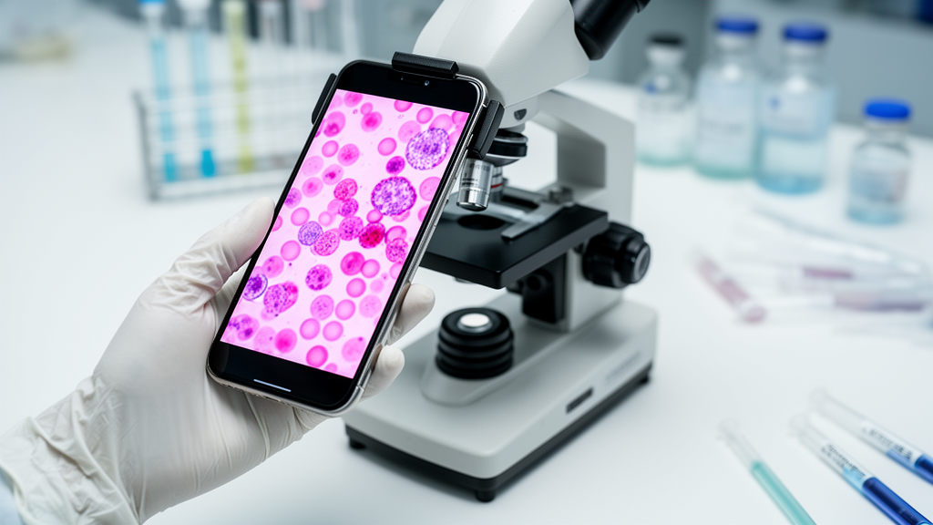
Abnormal vaginal discharge is one of the most common reasons women seek medical care, yet pinning down its cause remains surprisingly difficult in clinics around the world. The same symptoms — itching, discomfort, odor, unusual discharge — can signal vulvovaginal candidiasis, bacterial vaginosis, trichomoniasis, or gonococcal infection, each demanding a completely different treatment. Now a team of researchers at Chiang Mai University in Thailand has built an artificial intelligence system that can distinguish all of these conditions, plus healthy vaginal flora, from nothing more than a Gram-stained smear viewed through an ordinary light microscope and photographed with a smartphone held up to the eyepiece. The study, published in Frontiers in Artificial Intelligence, describes an explainable deep learning framework that not only reaches a diagnosis but shows clinicians exactly which microscopic structures justify it.
The innovation lies in how the system thinks about what it sees. Rather than forcing the AI to exhaustively find and label every object in a microscopic field, the researchers developed what they call detection-based image classification. The model scans each image for diagnostically relevant features — the filamentous pseudohyphae of Candida, the bacteria-coated clue cells of bacterial vaginosis, the flagellated trophozoites of Trichomonas vaginalis, or the Gram-negative diplococci of Neisseria gonorrhoeae tucked inside white blood cells. Finding even a single representative feature is enough to establish the image-level diagnosis, mirroring how experienced microbiologists actually work. This any-positive paradigm, conceptually related to multiple instance learning, means the system does not need pixel-perfect localization of every structure to be clinically useful.
To build the framework, the team assembled 1,966 Gram-stained microscopic images containing 11,069 annotated diagnostic objects across five categories: vulvovaginal candidiasis, bacterial vaginosis, trichomoniasis, gonococcal urethritis or cervicitis, and healthy Lactobacillus-dominated flora. Training slides came from archived teaching collections, while validation and test images were drawn from a completely separate set of prospectively collected patient specimens at Maharaj Nakorn Chiang Mai Hospital, ensuring no patient contributed to more than one dataset. Images were captured using a Samsung Galaxy S23 Ultra manually aligned with a conventional microscope eyepiece at 400x total magnification — no dedicated adapter, no digitized slide scanner, just the equipment already sitting in most clinical laboratories.
Three object detection architectures went head to head: YOLOv26, an anchor-free convolutional network; RT-DETR, an anchor-free vision transformer; and EfficientDet-D0, a lightweight anchor-based model. On the independent test set of 514 images, RT-DETR achieved the highest overall detection performance with a mean average precision at 50 percent overlap of 0.382, followed closely by YOLOv26 at 0.357, while EfficientDet-D0 lagged far behind at 0.116. Trichomoniasis and gonococcal infection were consistently the easiest classes to detect, with mAP50 values reaching 0.75, thanks to their distinctive morphology. Bacterial vaginosis, healthy flora, and candidiasis proved harder, largely because their diagnostic features are subtler and more morphologically heterogeneous — a finding that held even when the researchers tried balanced sampling and class-weighted losses to counteract uneven category representation.
Here is where the story takes a striking turn. Despite only modest box-level detection scores, the image-level diagnostic performance was exceptional. YOLOv26 achieved 95.72 percent accuracy with a macro F1-score of 0.9615, while RT-DETR reached 98.25 percent accuracy with an F1 of 0.9807 and a mean area under the curve of 0.9846. When predictions from multiple microscopic fields of the same specimen were aggregated by majority vote, RT-DETR correctly diagnosed all 96 unique test specimens — a perfect 100 percent at the specimen level. The gap between mediocre object localization and superb specimen-level diagnosis carries a broader lesson: conventional computer vision benchmarks like mAP50 may systematically underestimate the clinical value of AI microscopy systems whose real job is recognizing the presence of disease, not counting every object in the frame.
Interpretability was central to the design. Using Grad-CAM, EigenCAM, and Grad-CAM++ visualization techniques, the researchers showed that both models concentrated their attention on biologically plausible regions — clusters of diplococci within leukocyte-dense areas in gonococcal specimens, and clue-cell regions in bacterial vaginosis samples. Quantitative analysis of RT-DETR’s activation maps revealed a mean 2.40-fold enrichment of activation within annotated diagnostic regions relative to their share of the image, peaking at 3.96-fold for gonococcal infection. Eight independent clinicians and microbiologists then blindly rated the localization quality of the models’ bounding boxes, awarding Good or Excellent ratings in roughly 85 percent of valid evaluations for both architectures, with no statistically significant difference between them. Experts consistently preferred boxes that fully encompassed diagnostic structures — every single comparison involving complete pseudohypha coverage favored the more comprehensive box.
The efficiency benchmarks matter enormously for real-world deployment. YOLOv26 reached its best accuracy-efficiency balance at 800-pixel input resolution, matching RT-DETR’s peak detection accuracy while needing only 116.80 GFLOPs and 42.34 milliseconds per image on an NVIDIA T4 GPU. RT-DETR, by contrast, kept improving with resolution up to 1,280 pixels but demanded 421.73 GFLOPs and 169.04 milliseconds per image, along with 32.8 million parameters versus YOLOv26’s 21.8 million. For clinics in resource-limited settings where high-performance computing may be scarce, the lighter convolutional model offers a compelling trade-off, though cloud-based deployment could bring the full power of the transformer architecture to any connected device.
The clinical stakes are considerable. Microscopy remains a rapid, inexpensive diagnostic tool, yet studies have found it was performed in only 17.4 percent of visits among women with vulvovaginal symptoms, and accurate interpretation depends on experienced personnel subject to inter-observer variability. Untreated vaginal infections during pregnancy carry risks of adverse maternal and fetal outcomes, and misdiagnosis fuels unnecessary antimicrobial use. By pairing the microscopes and Gram stains already present in most laboratories with the smartphone already in most pockets, the framework could extend reliable differential diagnosis to settings that have never had access to specialized digital pathology infrastructure. Images with no detections above the confidence threshold are flagged as indeterminate and routed to manual review, a safeguard built into the proposed clinical workflow.
The researchers are careful to frame this as a proof-of-concept rather than a finished diagnostic product. The study was retrospective, single-institution, and used a standardized imaging workflow; the dataset contained no documented mixed infections, so the single-label framework may not capture coexisting conditions that occur in practice. Bounding-box annotations may underrepresent irregular structures like pseudohyphae, and clinical details such as pregnancy status and prior treatment were unavailable in the de-identified data. The authors call for rigorous prospective multi-center validation across varied microscopes, staining protocols, smartphone models, and patient populations before any autonomous diagnostic use. Still, the combination of high specimen-level accuracy, human-verifiable visual evidence, and hardware as ordinary as a phone and a light microscope makes this one of the most practical demonstrations yet that explainable AI can bring specialist-level microscopy to the front lines of global health.
Subject of Research: Explainable deep learning for smartphone-assisted microscopic differential diagnosis of vaginal discharge
Article Title: Interpretable detection-based image classification for differential diagnosis of vaginal discharge using smartphone-assisted microscopy
Article References: Sookkhee, S., Wongtapin, M., Phuriyaphan, T., Tonpiriyaphong, K., Wachirapaet, N., Wasanapitanont, S., Joysa, J., Pongpom, M., Lawan, N., Upaphong, P., & Wangsanut, T. (2026). Interpretable detection-based image classification for differential diagnosis of vaginal discharge using smartphone-assisted microscopy. Frontiers in Artificial Intelligence, 9, Article 1985517. https://doi.org/10.3389/frai.2026.1985517
Image Credits: AI Generated
DOI: 10.3389/frai.2026.1985517
Keywords: artificial intelligence, deep learning, medical imaging, vaginal discharge, bacterial vaginosis, vulvovaginal candidiasis, trichomoniasis, gonorrhea, smartphone microscopy, explainable AI, object detection, point-of-care diagnostics
Cite Scienmag News
Kristina Jarvis. (October 17, 2026). AI Turns a Smartphone and Microscope Into a Vaginal Infection Diagnostician. Scienmag. https://scienmag.com/ai-turns-a-smartphone-and-microscope-into-a-vaginal-infection-diagnostician/
Kristina Jarvis. "AI Turns a Smartphone and Microscope Into a Vaginal Infection Diagnostician." Scienmag, 17 October 2026, https://scienmag.com/ai-turns-a-smartphone-and-microscope-into-a-vaginal-infection-diagnostician/. Accessed 17 October 2026.
Kristina Jarvis. "AI Turns a Smartphone and Microscope Into a Vaginal Infection Diagnostician." Scienmag. October 17, 2026. https://scienmag.com/ai-turns-a-smartphone-and-microscope-into-a-vaginal-infection-diagnostician/


