


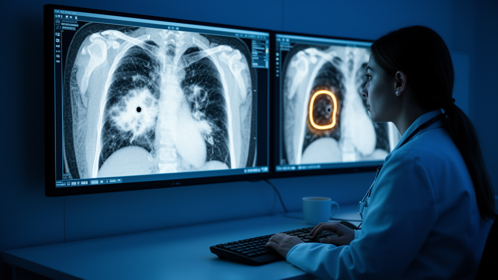
One of the most treacherous diagnostic puzzles in thoracic medicine has just been handed a powerful new tool. A team of researchers in China has developed an interpretable deep learning model that can distinguish a subtle, pneumonia-mimicking form of peripheral lung cancer from genuine inflammatory lung disease on contrast-enhanced CT scans, with performance that substantially exceeds conventional radiomics approaches. The multicenter retrospective study, published in BMC Cancer, offers a glimpse of how artificial intelligence could soon stand guard over one of radiology’s most consequential blind spots, potentially sparing patients from delayed cancer diagnoses that cost precious months of treatable time.
The clinical problem at the heart of the research is deceptively simple to state and maddeningly difficult to solve. Pneumonia-mimicking peripheral lung cancer, often abbreviated PLC, refers to lung tumors that arise in the outer regions of the lung and present on imaging as patchy opacities, ground-glass changes, or consolidation patterns that look almost indistinguishable from an infectious or inflammatory process. Radiologists confronting such lesions must weigh the possibility of malignancy against the far more common explanation of infection, and the stakes of that judgment are enormous. A false reassurance can allow an early-stage cancer to progress unchecked, while an unnecessary invasive workup exposes patients to procedural risk and anxiety. Conventional radiomics, which extracts hand-engineered quantitative features from medical images, has shown only limited diagnostic performance in this setting, leaving a genuine unmet need.
To close that gap, the research team assembled a dataset of 302 patients drawn from two medical centers, creating a multicenter cohort that lends the findings a degree of external validity often missing from single-institution machine learning studies. All patients had undergone venous-phase contrast-enhanced CT, the workhorse imaging protocol in which iodinated contrast agent highlights vascular and tissue enhancement patterns. Rather than feeding the network a single two-dimensional slice or the entire three-dimensional volume, the investigators adopted a middle path: they generated a 2.5D dataset by extracting seven consecutive slices centered on the largest axial cross-section of each lesion. This strategy captures contextual information above and below the most informative plane while keeping the data volume manageable, a pragmatic compromise between the simplicity of 2D analysis and the computational and data-hungry demands of full 3D modeling.
With the 2.5D data in hand, the team trained multiple deep learning architectures and compared their ability to separate cancer from mimicking inflammation. Among the candidates, ResNet101, a deep convolutional network whose residual connections allow very deep stacks of layers to be trained stably, emerged as the strongest performer, achieving an area under the receiver operating characteristic curve, or AUC, of 0.809 in the independent testing cohort, with a 95 percent confidence interval spanning 0.717 to 0.900. That figure, while respectable for a purely image-driven model, was only the beginning. The researchers then layered on more sophisticated machinery: multi-instance learning, a framework that treats each patient as a bag of image instances and learns from the collective evidence, and ensemble-based fusion strategies that combine the strengths of multiple models rather than betting on a single architecture.
The decisive leap in performance came when the deep learning features were fused with complementary sources of information. The team integrated predictive likelihood histogram features and bag-of-words features, techniques borrowed from natural language processing that summarize distributions of model outputs and image descriptors, with classical radiomics features and clinical variables. Statistical analysis identified three independent clinical predictors: patient sex, venous-phase CT attenuation values, and serum levels of carcinoembryonic antigen, a tumor marker frequently elevated in lung malignancies. When all of these strands were woven into a single combined model, the AUC rose to 0.874 in the training cohort and 0.855 in the independent testing cohort, the latter with a confidence interval of 0.774 to 0.936. In practical terms, the fused model correctly ranked cancerous lesions as more suspicious than their pneumonia lookalikes in roughly 85 to 87 percent of pairwise comparisons, a meaningful improvement over the image-only baseline.
Performance metrics alone, however, tell only part of the story, and the researchers were careful to interrogate their model with the full battery of validation tools now expected in clinical machine learning. Calibration curves demonstrated good agreement between predicted probabilities and observed outcomes, meaning that when the model expressed, say, a 70 percent likelihood of malignancy, roughly seven in ten such patients truly had cancer. Decision curve analysis, which quantifies the net clinical benefit of acting on a model’s predictions across a range of risk thresholds, showed that the combined model delivered higher net benefit than default strategies of treating all lesions as benign or all as malignant. These analyses matter because a model can achieve a dazzling AUC while being systematically overconfident or clinically useless at the thresholds where decisions are actually made, and the study’s results suggest the fused model avoids both traps.
Perhaps the most forward-looking aspect of the work is its commitment to interpretability, an attribute that has become a prerequisite for clinical acceptance of artificial intelligence. The team deployed two complementary explanation techniques. SHAP, or SHapley Additive exPlanations, draws on cooperative game theory to assign each input feature a quantified contribution to every individual prediction, revealing which clinical and imaging variables drove the model’s judgment in each case. Grad-CAM, or gradient-weighted class activation mapping, produces spatial heatmaps that highlight the image regions a convolutional network attended to when rendering its verdict. Together, these methods allow a radiologist to see not just what the model concluded but why, transforming an opaque algorithmic output into an auditable piece of diagnostic evidence that can be checked against human expertise.
The implications for clinical practice are considerable. A validated model of this kind could function as a silent second reader, flagging pneumonia-like lesions that warrant closer surveillance, PET imaging, or biopsy despite an ostensibly infectious appearance. Because the model relies on venous-phase contrast-enhanced CT, a scan already routinely obtained in the workup of indeterminate pulmonary lesions, it requires no new imaging protocol, no additional radiation exposure, and no extra cost beyond computation. The multicenter design, spanning the Second Affiliated Hospital of Nanchang University and the First Affiliated Hospital of Gannan Medical University, provides early reassurance that the model’s performance is not an artifact of one institution’s scanner fleet or patient mix, though prospective validation across broader and more diverse populations remains the essential next step before deployment.
The study also carries lessons for the wider field of medical artificial intelligence. Its architecture of success, combining a carefully engineered 2.5D input representation, multi-instance learning, ensemble fusion, and the deliberate integration of clinical laboratory data with imaging-derived features, illustrates that the best diagnostic models rarely come from raw images alone. The independent predictive value of carcinoembryonic antigen and venous-phase attenuation values underscores that biology and image intensity carry complementary signals, and that hybrid models can capture nuances invisible to either data stream in isolation. As screening programs expand and CT scans proliferate, tools that squeeze more diagnostic certainty from existing examinations will only grow in importance.
For now, the model remains a research instrument, its predictions confined to retrospective data and its authors appropriately cautious about immediate clinical use. Yet the trajectory is unmistakable. The work, supported by the Jiangxi Provincial Department of Science and Technology and conducted under the Declaration of Helsinki with ethics approval from Nanchang University, demonstrates that the pneumonia-mimicking lung cancer problem, long a source of diagnostic dread, is tractable to modern machine learning when the right data, architectures, and interpretability safeguards are brought to bear. If prospective studies confirm these results, the day may not be far off when every indeterminate, pneumonia-like spot on a chest CT is quietly accompanied by an algorithmic second opinion, one that has already learned to see through the disguise.
Subject of Research: Deep learning-based prediction of pneumonia-mimicking peripheral lung cancer on contrast-enhanced CT
Article Title: The predictive value of 2.5D deep learning model based on contrast-enhanced CT for pneumonia-mimicking peripheral lung cancer
Article References: Huang, H., Li, Q., Wang, J., Fu, X., Liu, Q., Liu, S., & Zuo, M. (2026). The predictive value of 2.5D deep learning model based on contrast-enhanced CT for pneumonia-mimicking peripheral lung cancer. BMC Cancer. https://doi.org/10.1186/s12885-026-17080-1
Image Credits: AI Generated
DOI: 10.1186/s12885-026-17080-1
Keywords: peripheral lung cancer, pneumonia-mimicking lung cancer, deep learning, 2.5D model, contrast-enhanced CT, radiomics, multi-instance learning, ensemble learning, ResNet101, SHAP, Grad-CAM, machine learning
Cite Scienmag News
Nathaniel Bowman. (October 8, 2026). AI Spots Lung Cancer Hiding Behind a Veil of Pneumonia on CT Scans. Scienmag. https://scienmag.com/ai-spots-lung-cancer-hiding-behind-a-veil-of-pneumonia-on-ct-scans/
Nathaniel Bowman. "AI Spots Lung Cancer Hiding Behind a Veil of Pneumonia on CT Scans." Scienmag, 8 October 2026, https://scienmag.com/ai-spots-lung-cancer-hiding-behind-a-veil-of-pneumonia-on-ct-scans/. Accessed 8 October 2026.
Nathaniel Bowman. "AI Spots Lung Cancer Hiding Behind a Veil of Pneumonia on CT Scans." Scienmag. October 8, 2026. https://scienmag.com/ai-spots-lung-cancer-hiding-behind-a-veil-of-pneumonia-on-ct-scans/

