


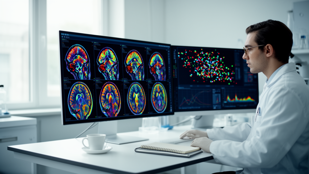
Artificial intelligence has moved from the margins of neuroradiology to its center, and a new systematic review now offers one of the clearest maps yet of where the technology actually stands. Published in Frontiers in Human Neuroscience, the review combed through more than a thousand records and distilled them into 123 peer-reviewed studies published between January 2020 and June 2026, all focused on a single question: how well can machine learning systems read brain images and help diagnose neurological disease? The answer, according to the authors, is both exhilarating and sobering. The field has surged forward in architectural sophistication, but its clinical maturity remains uneven, and the path to dependable deployment runs through data governance, transparent reporting, and independent multicenter testing rather than through bigger models alone.
The review followed PRISMA 2020 guidelines, the international standard for systematic evidence synthesis, and searched eight information sources using database-specific terms covering artificial intelligence, neuroimaging, neurological disease, and diagnostic analysis. The numbers tell a story of aggressive growth and rigorous pruning. The search initially identified 1,078 records; after removing 236 duplicates, 842 unique records were screened, 258 full texts were assessed in detail, and 123 studies survived a record-level eligibility re-audit. That funnel shape is familiar to anyone who follows the AI literature, but it underscores a point the authors make explicitly: enthusiasm for the topic far outstrips the number of studies that meet methodological standards for inclusion.
Once the evidence was assembled, the review mapped it across modalities, diseases, architectures, and validation practices. Magnetic resonance imaging dominated the included studies, appearing both as a primary imaging source and as auxiliary mapping data. Computed tomography was common in the acute settings where speed matters most, particularly stroke and intracranial hemorrhage, where every minute of delayed diagnosis translates into lost brain tissue. Positron emission tomography supported a narrower set of studies focused on molecular imaging and differential diagnosis, the delicate task of distinguishing between conditions that can look similar on structural scans. Functional MRI and diffusion imaging extended the picture into network-level and white-matter analysis, while electroencephalography carried the review’s scope beyond anatomy altogether, into the neurophysiological diagnosis of conditions such as epilepsy.
On the algorithmic side, convolutional neural networks and their three-dimensional variants remained the workhorses of the field, a reflection of how well their inductive biases suit volumetric medical images. But the review documents a clear generational shift in the more recent literature. Attention mechanisms, Transformer architectures, graph-based networks, self-supervised learning, generative models, multimodal approaches, and foundation models all appeared in selected recent studies, signaling a move away from narrow, single-task pattern recognition toward systems that can integrate diverse data streams and adapt to new tasks with less labeled data. This evolution mirrors broader trends in machine learning, but its consequences for clinical decision support are only beginning to be understood.
The diseases most frequently represented were stroke, dementia, and brain tumors, three conditions where imaging already plays a decisive clinical role and where the incentive to automate interpretation is correspondingly strong. Yet the authors are careful to resist the temptation to declare winners. Heterogeneity in patient populations, outcome definitions, and validation practices was so pronounced that the review explicitly declined to rank diseases, modalities, architectures, or validation strategies. Where meta-analysis would normally settle such questions, the sheer diversity of the evidence forced a narrative synthesis and descriptive mapping instead, a methodologically honest choice that many readers may find more useful than a spurious league table.
That heterogeneity is not a trivial complaint. When one study reports accuracy on a curated single-center dataset and another reports performance on a prospective multicenter cohort, the numbers are not directly comparable, and the review makes clear that incomplete validation extraction further muddied the picture. Many studies did not report the details that clinicians would need to judge readiness: whether the test set was truly independent, whether the model was calibrated, whether performance was stratified by demographic subgroup, and whether the system was evaluated in the messy context of an actual clinical workflow rather than on a static archive of images. These gaps, the authors argue, are the real bottleneck between laboratory performance and bedside reliability.
The review’s central conclusion is nuanced in a way that resists headline simplification, but its essence is this: the evidence suggests a movement from task-specific pattern recognition toward multimodal and context-rich decision support in selected settings, but it does not establish uniform methodological superiority or clinical maturity. In other words, the direction of travel is clear, and some destinations have been reached, but the field as a whole has not arrived. A model that excels at detecting hemorrhage on a curated CT dataset may still stumble on the scanner at the community hospital down the road, where acquisition protocols, patient demographics, and image quality differ in ways the training data never anticipated.
What would dependable translation actually require? The authors lay out a checklist that reads like a manifesto for the next phase of the field. Patient-level data governance, ensuring that privacy protections and consent frameworks keep pace with model development. Transparent reporting, so that others can reproduce and scrutinize results. Uncertainty estimation, so that a system can signal when it is unsure rather than confidently wrong. Independent multicenter testing, the gold standard for demonstrating that performance generalizes beyond the institution where the model was built. Prospective workflow evaluation, meaning that systems are assessed not just on archived images but in the live clinical environment where they will actually be used. And clinically meaningful endpoints, so that success is measured in patient outcomes rather than in area-under-the-curve statistics that may or may not translate into better care.
Each of these requirements addresses a documented failure mode. Models trained on data from a handful of centers have repeatedly been shown to latch onto site-specific artifacts rather than disease biology, a problem that only multicenter validation can expose. Systems that report a single headline accuracy metric can hide catastrophic performance on underrepresented subgroups, a problem that only stratified reporting can reveal. And tools evaluated only on retrospective archives can fail in ways no offline benchmark predicts, because the clinical workflow includes time pressure, competing information, and the judgment of the clinicians who must decide whether to trust the machine. The review’s insistence on prospective workflow evaluation is perhaps its most consequential recommendation, because it shifts the standard of evidence from what a model can do in principle to what it can do in practice.
For readers wondering whether AI will soon read their brain scans, the review offers a grounded answer: in selected settings, for selected tasks, elements of that future are already here, and the trajectory points toward increasingly multimodal, context-aware systems that fuse imaging with clinical data, genetics, and longitudinal history. But the same evidence makes clear that the bottleneck is no longer architectural imagination. The field has models to spare. What it needs now is the unglamorous infrastructure of trustworthy medicine: governed data, honest reporting, calibrated uncertainty, and trials that measure what actually matters to patients. The most important advances in AI for neurological diagnosis over the coming years may be less about novel networks and more about the discipline of proving, in the real world, that the networks we already have can be trusted with the human brain.
Subject of Research: Artificial intelligence methods for brain imaging and computer-aided diagnosis of neurological diseases
Article Title: Advances in artificial intelligence for brain imaging and computer-aided diagnosis of neurological diseases: a systematic literature review
Article References: Omarov, B. (2026). Advances in artificial intelligence for brain imaging and computer-aided diagnosis of neurological diseases: a systematic literature review. Frontiers in Human Neuroscience, 20, Article 1963855. https://doi.org/10.3389/fnhum.2026.1963855
Image Credits: AI Generated
DOI: 10.3389/fnhum.2026.1963855
Keywords: artificial intelligence, brain imaging, neurological diseases, computer-aided diagnosis, MRI, stroke, dementia, brain tumors, deep learning, systematic review, clinical validation, neuroimaging
Cite Scienmag News
Cassandra Pierce. (October 17, 2026). AI Reads the Brain: Systematic Review Maps the State of Neuroimaging Diagnosis. Scienmag. https://scienmag.com/ai-reads-the-brain-systematic-review-maps-the-state-of-neuroimaging-diagnosis/
Cassandra Pierce. "AI Reads the Brain: Systematic Review Maps the State of Neuroimaging Diagnosis." Scienmag, 17 October 2026, https://scienmag.com/ai-reads-the-brain-systematic-review-maps-the-state-of-neuroimaging-diagnosis/. Accessed 17 October 2026.
Cassandra Pierce. "AI Reads the Brain: Systematic Review Maps the State of Neuroimaging Diagnosis." Scienmag. October 17, 2026. https://scienmag.com/ai-reads-the-brain-systematic-review-maps-the-state-of-neuroimaging-diagnosis/


