


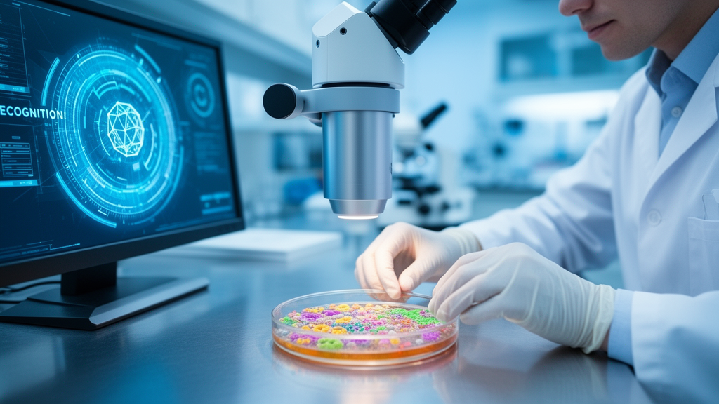
Reading an agar plate sounds simple: look for growth, describe the colonies, decide what to test next. Writing in Frontiers in Cellular and Infection Microbiology, a team led by Lukas Weber and Adrian Egli of the University of Zurich, with colleagues in Ethiopia, India, Bern and at ETH Zurich, surveys the state of automated image-based species identification and finds that deep learning models can already match or exceed human performance on several well-defined tasks, provided the results are interpreted within the narrow conditions in which they were validated.
The clinical problem the review addresses is longstanding. Culturing patient specimens takes 18 to 24 hours for most mesophilic bacteria, up to 96 hours for anaerobes, and weeks for slow growers such as Mycobacterium tuberculosis. Once colonies appear, technicians judge size, shape, colour and haemolysis, but morphology gives only a presumptive answer; a grey-greenish, crater-like colony with alpha haemolysis suggests Streptococcus pneumoniae, yet confirmation requires biochemical profiling or matrix-assisted laser desorption ionization time-of-flight mass spectrometry (MALDI-TOF MS). MALDI-TOF has largely replaced biochemical identification in high-income laboratories because it is fast and accurate, but its cost and infrastructure demands keep it out of reach for most low-resource settings. Plate reading itself is tedious, vulnerable to fatigue, and varies between observers.
The first deep learning milestone came from Huang and Wu, who trained a convolutional neural network (CNN) to classify colonies from 18 bacterial species using 4,982 images. The model reached an overall species-level classification accuracy of 0.967, but with a mean sensitivity of only 0.662 and precision of 0.723, the authors framed it as a pre-screening tool to narrow candidates before confirmatory tests. Around the same time, Majchrowska and colleagues released AGAR, an open dataset of 18,000 plate photographs covering single and mixed cultures of five microbial species with more than 336,000 annotated colonies, demonstrating that object detectors such as Faster R-CNN could localise and identify colonies simultaneously, a capability that matters for plates carrying mixed flora.
Growth detection, the simpler half of the problem, is essentially solved. A 2019 study of 1,581 urine cultures reported a sensitivity of 99.8 percent and specificity of 68.5 percent, and among 170 cases the automation flagged as positive but humans called negative, 116 involved microcolonies that technologists had missed. A larger 2020 study of 12,931 urine cultures confirmed 99.8 percent sensitivity with 72.0 percent specificity, with most discrepancies traced to expert rules classifying specimens as contamination rather than to threshold failures. For chromogenic screening tasks the numbers are similarly striking: the WASPLab Chromogenic Detection Module agreed with manual technologist readings in 90.1 percent of 104,730 vancomycin-resistant enterococci specimens, achieving 100 percent sensitivity and 89.5 percent specificity, and a combination of CHROMID Strepto B agar with PhenoMATRIX software detected group B streptococcus with 100 percent sensitivity after 48 hours, though specificity fell to 63.0 percent compared with 97.7 percent for nucleic acid amplification testing.
Species-level identification has progressed in step. Signoroni and colleagues built DeepColony, a multi-network system that segments colonies with a U-Net, classifies them with a CNN covering 32 species, then uses a Siamese network to cluster colonies by morphological similarity so that identical morphotypes are recognised as one organism rather than several spurious predictions. Evaluated on more than 5,000 clinical urine plates, DeepColony achieved roughly 90.6 percent top-1 and 97.6 percent top-3 identification accuracy in the review’s summary, and in a validation on 2,623 urine cultures agreed with manual interpretation 95.4 percent of the time, including 99.2 percent concordance on truly no-growth plates. Its main weakness was mixed growth: agreement dropped to 77 percent on plates human microbiologists deemed contaminated, because the AI tended to overcall clinically insignificant mixtures as significant, a conservative error the authors consider preferable to missing a pathogen.
Fungi have joined the pipeline more recently. Pouris and colleagues developed FungID, a transfer-learned VGG16 classifier trained on just 269 images of ten fungal species on chromogenic agar, which achieved a mean accuracy of 89.7 percent across species, though with a sensitivity of 65.3 percent and species-specific AUC values ranging from 0.60 to 0.93. The Zurich group’s own MoldVision goes further: a smartphone-imaging workflow tested on 161 clinical isolates of Penicillium, Aspergillus flavus, Aspergillus fumigatus, Fusarium and Cladosporium, using more than 4,000 images collected over five days of growth. The best dual-head VGG16 model reached a mean AUROC of 92.7 percent with 68.7 percent sensitivity, and in mature cultures often matched or outperformed expert readers. Colony maturity proved decisive: the F1 score rose from 38.8 percent on days one to two to 92.1 percent on days three to five, a pattern the authors say highlights how biologically immature phenotypes resist classification.
These research tools are already embedded in commercial automation. Copan’s WASPLab and BD’s Kiestra systems combine robotic plate handling, automated incubators and digital imaging, and their software suites, PhenoMATRIX and Kastle, flag plates with no significant growth, count colony-forming units and recognise colony colour types. Clever Culture Systems’ APAS Independence became the first FDA-cleared AI-based plate reader in 2019, focused on urine cultures, and one report found roughly an 80 percent reduction in hands-on review of negative urine cultures when automation was paired with AI screening. One laboratory shortened incubation-to-report time for urine cultures by about four hours on average. Current commercial systems stop short of fully automated species identification, but the review notes they can already label pink colonies on validated chromogenic urine agar as Escherichia coli with high confidence, and typically combine learned models with rule-based expert logic, such as reporting a pure culture when a single morphotype exceeds 100 CFU.
Performance outside the training environment remains the central caveat. A whole-plate deep-learning classifier trained on 114,074 urine culture images from two institutions achieved within-institution AUCs of at least 0.98, but cross-institution performance dropped to at least 0.90, and the study’s authors cited subjective reference labels and non-standardised interpretation classes as limitations. Unusual lighting, unfamiliar agar types or species absent from training data can degrade models substantially, and under the European in vitro diagnostic regulation the review warns this could create commercial dependencies on specific plate manufacturers. Reporting heterogeneity compounds the problem: many studies cite only routine diagnostics as ground truth without specifying whether identification relied on biochemical tests, MALDI-TOF, PCR or sequencing, which hampers cross-study comparison. The review’s authors explicitly declined to pool the numerical results they surveyed, arguing that differences in task definition, datasets and validation strategies make a meta-analysis methodologically inappropriate.
The review also points beyond conventional cameras. Li and colleagues coupled a thin-film transistor image sensor with two neural networks, one detecting growth and one classifying Escherichia coli versus other coliforms, achieving a 97.3 percent CFU detection rate at nine hours and a 91.6 percent classification recovery rate at 12 hours, saving more than 12 hours compared with conventional methods. Paquin and colleagues reached a 96.0 percent average detection rate within eight hours using a lens-free CMOS approach. Elastic light scattering has classified colonies with accuracies of 95 to 100 percent in compact laser-based systems, and Schultz and colleagues distinguished Staphylococcus aureus from non-S. aureus isolates with a 94 percent correct identification rate after six hours of incubation. Hyperspectral imaging reached a mean accuracy of 95.06 percent for Shiga toxin-producing E. coli serotypes, and lens-free plus infrared imaging classified ten bacterial strains at 94.4 percent accuracy.
For the authors, the priority is no longer higher accuracy under controlled conditions but reliable, auditable performance across laboratories, imaging systems and complex real-world cultures. They argue AI should be deployed as a risk-stratified decision-support tool, safely auto-clearing true negatives while flagging ambiguous plates for human review, with expert oversight retained for pre-analytics, quality assurance and exception handling. Smartphone-based imaging paired with lightweight models such as those runnable on TensorFlow Lite could extend growth detection, colony enumeration and presumptive identification to laboratories without MALDI-TOF or full automation, though the review insists such tools must meet the same external validation, quality control and regulatory standards as high-end systems. Under current regulation, pre-trained locked models are considered more realistic than continuously retrained ones, and any software update would require renewed validation against standardised image panels. The technology, in short, is ready to augment microbiologists; the evidence base, the authors conclude, must now catch up.
Subject of Research: Artificial intelligence and deep learning for automated species identification of bacteria and fungi from digital images of culture plates in diagnostic microbiology.
Article Title: State-of-the-art in automated image-based species identification systems
Article References: Weber, L., Hussen, M. M., Veeraraghavan, B., Meijer, E., Gütlin, Y., Guerrero-López, A., Brüningk, S., Nolte, O., Jutzeler, C., & Egli, A. (2026). State-of-the-art in automated image-based species identification systems. Frontiers in Cellular and Infection Microbiology, 16, Article 1818023. https://doi.org/10.3389/fcimb.2026.1818023
Image Credits: AI Generated
DOI: 10.3389/fcimb.2026.1818023
Keywords: artificial intelligence, deep learning, convolutional neural networks, culture plates, microbial identification, diagnostic microbiology, MALDI-TOF MS, laboratory automation, smartphone imaging, fungi, urine cultures, clinical validation
Cite Scienmag News
Blake Davidson. (October 27, 2026). AI Reads Petri Dishes and Names the Microbes Faster Than Ever. Scienmag. https://scienmag.com/ai-reads-petri-dishes-and-names-the-microbes-faster-than-ever/
Blake Davidson. "AI Reads Petri Dishes and Names the Microbes Faster Than Ever." Scienmag, 27 October 2026, https://scienmag.com/ai-reads-petri-dishes-and-names-the-microbes-faster-than-ever/. Accessed 27 October 2026.
Blake Davidson. "AI Reads Petri Dishes and Names the Microbes Faster Than Ever." Scienmag. October 27, 2026. https://scienmag.com/ai-reads-petri-dishes-and-names-the-microbes-faster-than-ever/


