


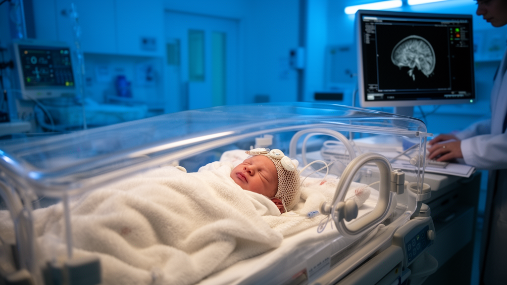
Every year, countless newborns experience birth asphyxia, a terrifying moment when oxygen flow to the baby is interrupted during delivery. For the infants who survive, doctors face an urgent question: did the brain suffer damage from the oxygen deprivation, a condition known as hypoxic-ischemic encephalopathy, or HIE? The answer shapes everything from intensive monitoring to follow-up care, and ultimately the child’s developmental trajectory. Yet one of the most frustrating scenarios in neonatal medicine is the baby whose conventional magnetic resonance imaging scan looks entirely normal to the radiologist’s eye, even though clinical suspicion of brain injury remains high. A new study published in BMC Medical Imaging suggests that machine learning can find the injury that human eyes miss, combining subtle mathematical signatures extracted from MRI scans with routine clinical data to detect HIE in term neonates whose images appear visually unremarkable.
The research, led by Yong-hao Yue, Shi-jin Zhong, Hui-min Mao, Yu-shan Chen, and Wan-liang Guo of the Radiology Department at the Children’s Hospital of Soochow University, together with neonatologist Xin Ding and radiologist Yong-qiang Ma of the Radiology Department of Yulin, tackles a well-known blind spot in neonatal imaging. Conventional MRI sequences, including T1-weighted, T2-weighted, and diffusion-weighted imaging, are the workhorses of neonatal brain assessment, but early or subtle hypoxic-ischemic injury can fall below the threshold of visual detection. Radiologists read images by looking for visible changes in signal intensity, tissue swelling, or patterns of injury in vulnerable brain regions. When those changes are faint or absent, the scan is reported as normal, and the infant may be classified as low risk even though the underlying biology tells a different story.
The technique the Chinese team deployed is called radiomics, a branch of medical imaging analysis that converts pictures into high-dimensional data. Rather than relying on what a human observer can see, radiomics algorithms extract hundreds of quantitative features from a defined region of interest: measures of texture, intensity distribution, shape, and the fine spatial relationships between neighboring pixels. These features capture tissue properties that are invisible to the naked eye but statistically distinguishable. In this study, the researchers focused on two regions of the newborn brain that are particularly vulnerable to oxygen deprivation: the basal ganglia and the thalami, deep gray matter structures that act as critical relay stations for movement control and sensory processing. Injury to these areas is a hallmark of severe HIE, but early changes can be extremely subtle.
To build their model, the team assembled a retrospective cohort of 297 term neonates with birth asphyxia treated between January 2020 and December 2025. The cohort was randomly divided into a training set and a test set in a seven-to-three ratio, a standard practice that allows the model to learn from one portion of the data while being evaluated on data it has never seen. Regions of interest were manually delineated within the basal ganglia and thalami on T1-weighted, T2-weighted, and diffusion-weighted images. From each region and each sequence, the researchers extracted radiomics features and then screened them using established feature-reduction techniques, including minimum redundancy maximum relevance selection and the least absolute shrinkage and selection operator, known as LASSO, alongside random forest methods. This screening process distilled the raw feature pool into six sequence- and region-specific radiomics scores, each a compact numerical summary of the hidden imaging signature in a particular brain area and MRI sequence.
The researchers then built five competing models to determine the best strategy. The first was a purely clinical model, built from clinical predictors identified through univariate and multivariate logistic regression, drawing on variables such as perinatal history and laboratory findings. The second and third were single-region radiomics models, one based on the basal ganglia and one on the thalami. The fourth was a radiomics fusion model that pooled information across regions and sequences. The fifth, and ultimately the winner, was a combined model that integrated the clinical predictors with the radiomics features. Performance was evaluated rigorously using receiver operating characteristic analysis, with the area under the curve, or AUC, serving as the headline metric, and the DeLong test used to determine whether differences between models were statistically significant.
The results were striking. The combined model achieved an AUC of 0.958 in the training set, 0.915 in the test set, and, most importantly, 0.878 in a prospective validation set of 54 neonates recruited from the Children’s Hospital of Soochow University and Yulin Children’s Hospital between January and April 2026. An AUC of 0.5 would indicate performance no better than a coin flip, while 1.0 represents perfect discrimination; values approaching 0.9 in an independent prospective cohort represent a level of accuracy that few clinical prediction tools in neonatology achieve. The DeLong test confirmed that the combined model significantly outperformed all of the comparator models, including the radiomics-only fusion model, demonstrating that clinical context and quantitative imaging information each contribute unique diagnostic value that neither provides alone.
Accuracy alone, however, is not enough for a tool intended for clinical use. A prediction model must also be calibrated, meaning that when it says a baby has a 70 percent chance of having HIE, roughly 70 percent of such babies should actually have the condition. The researchers assessed this using calibration curves and the Hosmer-Lemeshow framework, finding acceptable agreement between predicted and observed outcomes. They also performed decision curve analysis, a method that quantifies the net clinical benefit of acting on the model’s predictions across a range of decision thresholds, and found that the combined model offered a potential net benefit compared with treating all infants or treating none. Together, these analyses suggest the tool is not merely statistically impressive but potentially actionable at the bedside.
One of the most forward-looking elements of the study is its use of SHAP analysis, short for SHapley Additive exPlanations, a technique borrowed from game theory that decomposes each individual prediction to reveal how much every input variable contributed to it. In an era when clinicians are rightly wary of black-box algorithms, SHAP provides a window into the model’s reasoning. In this study, the interpretability analysis indicated that both clinical variables and radiomics features contributed meaningfully to the predictions, with features derived from T2-weighted and diffusion-weighted imaging exhibiting the highest importance. This makes biological sense: T2-weighted sequences are sensitive to tissue water content and edema, while diffusion-weighted imaging detects restrictions in the microscopic movement of water molecules, one of the earliest cellular consequences of hypoxic-ischemic injury. The model, in effect, learned to read the same physiological processes that radiologists look for, but at a resolution far finer than human perception allows.
The clinical implications are significant. For neonates with birth asphyxia and visually negative MRI findings, the current standard of care relies on clinical judgment, laboratory markers, and serial imaging, with the risk that genuinely injured infants are under-triaged and miss the window for interventions such as follow-up neuroprotective strategies and early developmental support. An objective, quantitative tool that flags hidden injury could help clinicians stratify risk more precisely, guide the intensity of monitoring, and determine which infants need closer neurodevelopmental follow-up. The authors emphasize that the combined model is intended to complement, not replace, routine clinical assessment, serving as an additional layer of evidence in a population where every piece of information matters.
Caveats remain, as they do with any emerging technology. The retrospective portion of the cohort, while large, reflects the patient population and imaging protocols of the participating hospitals, and the prospective validation set, though crucial, included 54 infants from two centers. Broader multi-center validation across diverse scanners, sequences, and populations will be needed before such a model becomes standard practice. The study also received support from the Natural Science Foundation of Jiangsu Province, the National Natural Science Foundation of China, and the Suzhou Medical Key Discipline program, reflecting the growing institutional investment in medical artificial intelligence. Still, the trajectory is clear: the fusion of deep clinical knowledge with quantitative imaging analysis is steadily converting the radiologist’s intuition into reproducible numbers. For the smallest and most vulnerable patients, those numbers may soon mean the difference between an injury caught in time and one discovered too late.
Subject of Research: A clinical-radiomics machine learning model for identifying hypoxic-ischemic encephalopathy in term neonates with birth asphyxia and visually normal MRI findings
Article Title: Beyond visually negative MRI: a clinical–radiomics combined model for identifying hypoxic–ischemic encephalopathy in term neonates with birth asphyxia
Article References: Yue, Y.-H., Zhong, S.-J., Mao, H.-M., Chen, Y.-S., Ding, X., Ma, Y.-Q., & Guo, W.-L. (2026). Beyond visually negative MRI: a clinical–radiomics combined model for identifying hypoxic–ischemic encephalopathy in term neonates with birth asphyxia. BMC Medical Imaging. https://doi.org/10.1186/s12880-026-02764-8
Image Credits: AI Generated
DOI: 10.1186/s12880-026-02764-8
Keywords: hypoxic-ischemic encephalopathy, birth asphyxia, neonates, MRI, radiomics, machine learning, basal ganglia, thalami, diffusion-weighted imaging, SHAP, decision curve analysis, BMC Medical Imaging
Cite Scienmag News
Ophelia Keating. (October 17, 2026). AI Reads Hidden Brain Injury in Newborns When MRI Looks Normal. Scienmag. https://scienmag.com/ai-reads-hidden-brain-injury-in-newborns-when-mri-looks-normal/
Ophelia Keating. "AI Reads Hidden Brain Injury in Newborns When MRI Looks Normal." Scienmag, 17 October 2026, https://scienmag.com/ai-reads-hidden-brain-injury-in-newborns-when-mri-looks-normal/. Accessed 17 October 2026.
Ophelia Keating. "AI Reads Hidden Brain Injury in Newborns When MRI Looks Normal." Scienmag. October 17, 2026. https://scienmag.com/ai-reads-hidden-brain-injury-in-newborns-when-mri-looks-normal/


