


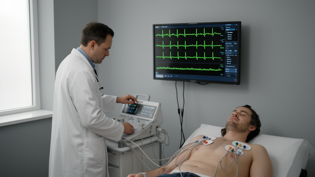
Artificial intelligence has been reading electrocardiograms for years, and the results have often sounded extraordinary: algorithms that spot heart attacks human eyes miss, that predict atrial fibrillation before it happens, and that detect failing hearts from a routine 10-second tracing. But a sweeping new critical review argues that the field’s headline-grabbing accuracy scores conceal a far messier reality — one in which some AI-ECG applications are already reshaping emergency care while others remain stuck at the stage of promising but unproven laboratory exercises.
The review, published in Frontiers in Cardiovascular Medicine, was led by Huanhuan Chen of the University of Shanghai for Science and Technology together with Jingjuan Huang and Xumin Hou of Shanghai Chest Hospital, Shanghai Jiao Tong University. Rather than cataloguing model architectures or celebrating benchmark scores, the team set out to answer a deceptively simple question: which AI-ECG applications are actually ready for clinical use, and which are not? To do so, they searched PubMed, IEEE Xplore, and Web of Science from January 2022 through July 2026, adding earlier landmark studies through backward citation tracking. Seventy-seven publications ultimately informed the synthesis.
The authors appraised each application against five clinically oriented criteria: whether the intended use is well defined, whether the reference standard — the ground truth used to train and test the model — is reliable, whether the model has been externally validated across populations and devices, whether its outputs are clinically actionable, and whether there is evidence that the model improves workflow or patient outcomes. This framework deliberately separates technical performance from translational maturity, a distinction the authors argue has been routinely blurred in the literature.
The clearest success stories involve ST-segment elevation myocardial infarction (STEMI) triage and screening for reduced left ventricular ejection fraction. In a pragmatic randomized trial published in NEJM AI, an AI-ECG algorithm for rapid STEMI identification achieved a positive predictive value of 89.5 percent and a negative predictive value of 99.9 percent, and AI-assisted triage measurably shortened door-to-balloon and ECG-to-balloon times — the kind of workflow-level evidence that retrospective accuracy studies cannot provide. Similarly, a randomized controlled trial of an AI-ECG alert for reduced ejection fraction among non-cardiology inpatients, published in BMC Medicine, increased new diagnoses of reduced LVEF, raised the positive predictive value of subsequent echocardiography from 20.2 to 34.2 percent, and boosted cardiology consultation rates from 23.5 to 29.3 percent — all without increasing overall echocardiography use.
Acute myocardial infarction rule-out has also advanced, reaching prospective multicenter diagnostic validation. In the ROMIAE study, a ResNet-based model using only the initial 12-lead ECG achieved an area under the receiver operating characteristic curve of 0.878, and at a predefined low-risk threshold it reached 99.6 percent sensitivity and 99.1 percent negative predictive value. Hypertrophic cardiomyopathy screening has progressed through international external validation — with an AUC of 0.922 across cohorts from Switzerland, the United Kingdom, and South Korea — and through prospective implementation: an FDA-cleared alert system screened 145,848 patients across five healthcare systems, generated 4,348 alerts, and identified 17 new HCM diagnoses, with a median of 7.5 days from ECG to diagnostic imaging.
But the picture darkens considerably elsewhere. Detection of occlusion myocardial infarction — a condition the review notes is missed by conventional ST-elevation criteria, which show a pooled sensitivity of only 43.6 percent for detecting acute coronary occlusion — remains at the external diagnostic-validation stage. Deep learning models such as ECG-SMART-NET have achieved impressive AUCs around 0.95 using angiographically confirmed occlusion as the reference standard, yet no prospective interventional study has tested whether AI-guided catheterization-laboratory activation actually improves reperfusion decisions without triggering excessive false activations. Prediction of occult or future atrial fibrillation from sinus-rhythm ECGs, including a large Veterans Affairs study of more than 900,000 outpatient ECGs that achieved an AUROC of 0.93, remains a risk-enrichment strategy rather than a diagnostic tool, partly because the reference standard depends on subsequent clinical detection of AF.
Valvular heart disease, heart failure with preserved ejection fraction, long-term heart-failure prediction, and cardiac amyloidosis screening all remain primarily validation-stage or risk-enrichment applications. For valvular disease, models can detect moderate-to-severe aortic stenosis, mitral regurgitation, and other lesions with AUCs generally between 0.77 and 0.88, but positive predictive value is heavily dependent on disease prevalence, and no prospective workflow evidence exists. For cardiac amyloidosis, the Mayo Clinic AI-ECG model achieved an AUC of 0.84, but performance dropped substantially in Hispanic patients and in ECGs with left ventricular hypertrophy or left bundle branch block — a pattern the authors flag as a transportability and equity concern. For heart failure with preserved ejection fraction, the lack of a single definitive reference standard makes the prediction target inherently unstable.
Underlying all of these gaps, the review identifies a consistent set of constraints: label quality, calibration, transportability across devices and institutions, operating-threshold selection, and the near-universal absence of patient-outcome evidence. The authors emphasize that a high AUROC says nothing about how a model behaves at the specific threshold where it will actually be deployed, how many false positives it will generate in a low-prevalence screening population, or whether clinicians will act on its output. They also caution that multimodal models — those combining ECG with clinical variables, biomarkers, or imaging — may derive much of their apparent advantage from established non-ECG predictors rather than from genuinely novel electrical signatures.
The review’s bottom line is measured rather than dismissive. AI-ECG, the authors conclude, is best positioned as clinician-supervised decision support linked to explicit downstream testing and care pathways — not as an autonomous diagnostician. The applications that have progressed furthest are precisely those in which the model’s output connects to an established clinical action: urgent reperfusion for STEMI, echocardiography for reduced ejection fraction, imaging and genetic counseling for suspected hypertrophic cardiomyopathy. Where that connection is missing, even technically brilliant models remain solutions in search of a clinical problem. The next phase of the field, the authors argue, must shift from retrospective discrimination studies toward prospective, device-diverse, outcome-oriented trials that test whether AI-ECG actually changes what happens to patients — not just what appears on a receiver operating characteristic curve.
Subject of Research: Translational maturity of artificial intelligence-enabled electrocardiography across cardiovascular diseases
Article Title: Translational maturity of artificial intelligence-enabled electrocardiography varies across cardiovascular diseases
Article References: Chen, H., Huang, J., & Hou, X. (2026). Translational maturity of artificial intelligence-enabled electrocardiography varies across cardiovascular diseases. Frontiers in Cardiovascular Medicine, 13, Article 1940621. https://doi.org/10.3389/fcvm.2026.1940621
Image Credits: AI Generated
DOI: 10.3389/fcvm.2026.1940621
Keywords: AI-ECG, electrocardiography, STEMI, myocardial infarction, heart failure, atrial fibrillation, hypertrophic cardiomyopathy, cardiac amyloidosis, valvular heart disease, external validation, clinical decision support, translational medicine
Cite Scienmag News
Ophelia Keating. (October 16, 2026). AI-Reads ECGs: How Far Has the Hype Actually Traveled Into the Clinic? Scienmag. https://scienmag.com/ai-reads-ecgs-how-far-has-the-hype-actually-traveled-into-the-clinic/
Ophelia Keating. "AI-Reads ECGs: How Far Has the Hype Actually Traveled Into the Clinic?" Scienmag, 16 October 2026, https://scienmag.com/ai-reads-ecgs-how-far-has-the-hype-actually-traveled-into-the-clinic/. Accessed 16 October 2026.
Ophelia Keating. "AI-Reads ECGs: How Far Has the Hype Actually Traveled Into the Clinic?" Scienmag. October 16, 2026. https://scienmag.com/ai-reads-ecgs-how-far-has-the-hype-actually-traveled-into-the-clinic/


