


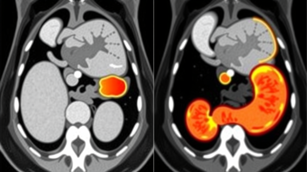
Gastric cancer remains one of the world’s most formidable malignancies, and for the majority of patients diagnosed with the disease, malnutrition is an ever-present threat. Driven by systemic metabolic alterations and the punishing side effects of cancer therapy, this nutritional decline often manifests as sarcopenia, a progressive and generalized skeletal muscle disorder that serves as an independent predictor of adverse clinical outcomes. Identifying sarcopenia early is essential for optimizing risk stratification and enabling timely, individualized nutritional interventions. Yet the conventional diagnostic process—assessing muscle strength, muscle quantity or quality, and physical performance—is labour-intensive and time-consuming, limiting its application in routine clinical practice. Now, a team of researchers from Wenzhou Medical University has developed an artificial intelligence model that could change how clinicians screen for this condition, fusing deep learning, radiomics, and clinical data into a single automated assessment tool.
At the heart of the new approach is a multimodal fusion model that integrates three distinct but complementary data streams: clinical variables such as gender, age, body mass index, haemoglobin, and albumin; high-dimensional radiomics features extracted from computed tomography images; and deep learning features derived from a ResNet50 convolutional neural network. The research, published in the Journal of Cachexia, Sarcopenia and Muscle, drew on data from 1067 patients who underwent gastric cancer surgery at two hospitals affiliated with Wenzhou Medical University between 2014 and 2022. Of these, 978 patients formed the training and validation sets, while 89 patients from a second hospital served as an external test set to evaluate the model’s ability to generalize to unseen data.
Defining sarcopenia accurately was central to the study’s design. Rather than relying solely on the skeletal muscle index (SMI) measured at the third lumbar vertebral (L3) level—the widely accepted reference standard for assessing muscle mass—the researchers adhered to the European Working Group on Sarcopenia in Older People (EWGSOP2) criteria. Sarcopenia was defined by the presence of both low muscle strength and low muscle quantity or quality, while severe sarcopenia required the additional presence of low physical performance. Muscle strength was assessed using grip strength, with cutoff values defined as less than 28 kilograms for men and less than 18 kilograms for women. Physical performance was evaluated using the 6-meter gait speed test, with a speed below 1.0 meters per second indicating low performance. This comprehensive definition better captures the combined structural and functional nature of the condition compared with studies that rely on CT-derived muscle area alone.
The technical pipeline behind the model is as sophisticated as it is elegant. Automated skeletal muscle segmentation was performed using TotalSegmentator, a deep learning tool that eliminates the need for manual delineation by radiologists. Axial slices at the L3 level, along with one adjacent slice above and below, were extracted from preoperative abdominal CT scans. From these images, the researchers used the PyRadiomics library to extract 1288 quantitative features spanning shape, first-order intensity, and texture classes. After Z-score normalization and removal of highly correlated features, LASSO regression with 10-fold cross-validation whittled the set down to 26 sarcopenia-related features. An extreme gradient boosting (XGBoost) model trained on these features achieved an area under the curve (AUC) of 0.93 in the training set and 0.85 in the validation set, demonstrating the diagnostic power of radiomic texture information alone.
Alongside the radiomics approach, the team developed 2D and 2.5D deep learning models based on the ResNet50 architecture pretrained on the ImageNet dataset. The 2D model processed a single L3-level muscle slice, while the 2.5D model combined three consecutive slices as a three-channel input, capturing critical anatomical spatial continuity across adjacent sections. Transfer learning was performed on the training set, with network parameters iteratively updated using backpropagation and the Adam optimizer. Data augmentation strategies, including random horizontal and vertical flipping, helped improve generalizability. After training, 2048 deep learning features were extracted from the penultimate global average pooling layer of the network for each patient. The 2.5D model outperformed its 2D counterpart, achieving an AUC of 0.83 in the external test set compared with 0.81, prompting the team to select 2.5D features for the final multimodal fusion.
The integration of these heterogeneous data sources was accomplished through a transformer-based architecture that treats clinical variables, radiomics features, and deep learning features as three independent input modalities. Each modality was separately fed into modality-specific encoding networks composed of fully connected hidden layers to learn high-dimensional discriminative representations. Dropout regularization was applied during encoding to reduce overfitting. The encoded features from all three modalities were then projected into a 64-dimensional latent space and fused using a transformer encoder with two encoder layers and eight attention heads—a mechanism borrowed from natural language processing that allows the model to weigh the relative importance of different pieces of information dynamically. The model’s output consisted of two independent classification heads, one estimating the probability of sarcopenia and the other estimating the probability of severe sarcopenia.
The results are striking. In the external test set, the integrated model achieved an AUC of 0.89 for sarcopenia and 0.84 for severe sarcopenia, with accuracy rates of 0.80 and 0.73 respectively. In the training set, performance was even more impressive, with AUCs of 0.95 for sarcopenia and 0.93 for severe sarcopenia. Decision curve analysis confirmed that the model provided positive net benefit across clinically relevant threshold probabilities, suggesting genuine utility in real-world screening scenarios. Among the cohort, the prevalence of sarcopenia was 20.0 percent and severe sarcopenia was 12.3 percent, proportions that remained consistent across all three patient sets. Kaplan-Meier survival analysis revealed that patients with sarcopenia had significantly poorer overall survival than those without, reinforcing the clinical urgency of early identification.
Interpretability was not neglected in the rush toward higher performance. The researchers employed SHapley Additive exPlanations (SHAP) analysis to quantify the contribution of individual features in both the radiomics and XGBoost models. Notably, the leading SHAP features in the XGBoost model spanned all three modalities, and several were significantly correlated with grip strength and SMI, supporting the clinical and biological relevance of the features the model had learned to prioritize. This kind of transparency matters in a medical context, where clinicians need to trust that a model’s predictions are grounded in meaningful physiological signals rather than spurious correlations.
The implications extend beyond gastric cancer. Tumor-associated chronic inflammation disrupts protein metabolism and impairs a patient’s ability to tolerate surgical stress, making muscle wasting a reflection of underlying global physiological vulnerability rather than a simple reduction in body weight. As these pathophysiological alterations directly influence postoperative recovery and long-term prognosis, comprehensive risk stratification has become central to personalized prehabilitation. The automated workflow established by this study facilitates the integration of high-dimensional radiological data with clinical information and may provide a feasible approach for early screening, helping to identify high-risk patients who could benefit from earlier targeted nutritional and functional interventions before surgery even begins.
Despite the promising results, the researchers acknowledge several limitations. Further validation in large-scale prospective cohorts is needed to confirm the model’s real-world predictive performance, and incorporating volumetric data from a broader range of abdominal levels may provide a more comprehensive assessment of systemic muscle wasting. The study also relied primarily on CT-based assessments, so generalizability to clinical settings that use different body composition technologies—such as bioelectrical impedance analysis or dual-energy X-ray absorptiometry—remains to be established. Nevertheless, by simplifying and optimizing the evaluation workflow, the model reduces reliance on labour-intensive manual assessments and provides a foundation for earlier implementation of individualized nutritional and functional interventions in routine clinical practice, offering a glimpse of how artificial intelligence can quietly transform the care of some of medicine’s most vulnerable patients.
Subject of Research: A multimodal artificial intelligence model combining radiomics, deep learning, and clinical variables for automated detection of sarcopenia in gastric cancer patients.
Article Title: Integrated Assessment of Sarcopenia in Patients with Gastric Cancer Using Deep Learning and Radiomics
Article References: Zhi, H., Zheng, J., Chen, H., Yang, X., Jiang, Z., Shi, A., Wu, D., Lv, L., He, J., Chen, X., Pan, B., Shen, X., & Zhang, W. (2026). Integrated Assessment of Sarcopenia in Patients with Gastric Cancer Using Deep Learning and Radiomics. Journal of Cachexia, Sarcopenia and Muscle, 17(5), Article e70377. https://doi.org/10.1002/jcsm.70377
Image Credits: AI Generated
DOI: 10.1002/jcsm.70377
Keywords: sarcopenia, gastric cancer, deep learning, radiomics, computed tomography, multimodal fusion, transformer, muscle wasting, XGBoost, ResNet50, clinical prediction, nutritional intervention
Cite Scienmag News
Ophelia Keating. (September 21, 2026). AI Model Spots Muscle-Wasting Syndrome in Gastric Cancer Patients from Routine CT Scans. Scienmag. https://scienmag.com/ai-model-spots-muscle-wasting-syndrome-in-gastric-cancer-patients-from-routine-ct-scans/
Ophelia Keating. "AI Model Spots Muscle-Wasting Syndrome in Gastric Cancer Patients from Routine CT Scans." Scienmag, 21 September 2026, https://scienmag.com/ai-model-spots-muscle-wasting-syndrome-in-gastric-cancer-patients-from-routine-ct-scans/. Accessed 4 October 2026.
Ophelia Keating. "AI Model Spots Muscle-Wasting Syndrome in Gastric Cancer Patients from Routine CT Scans." Scienmag. September 21, 2026. https://scienmag.com/ai-model-spots-muscle-wasting-syndrome-in-gastric-cancer-patients-from-routine-ct-scans/


