


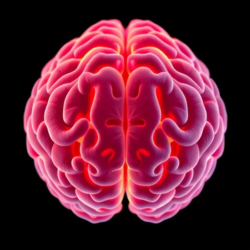
USC researchers have developed an artificial intelligence system that creates detailed maps showing how individual regions of the human brain appear to age. Unlike conventional “brain age” models, which compress a scan into a single number, the new deep-learning approach measures aging throughout the brain at the voxel level—the scale of the tiny three-dimensional units that make up magnetic resonance imaging data. The result is a regional portrait of brain aging that may reveal why some cognitive abilities remain resilient while others decline.
The model was developed by a team led by Andrei Irimia, associate professor at the USC Leonard Davis School of Gerontology. Researchers trained a deep neural network using MRI scans from 14,748 cognitively healthy adults between 19 and 100 years old. These scans came from six large public datasets, including the UK Biobank, the Human Connectome Project and the Alzheimer’s Disease Neuroimaging Initiative. By learning how healthy brain structures typically vary across adulthood, the system established a reference pattern against which individual scans could be compared.
When presented with a new MRI scan, the model estimates the apparent biological age of different brain locations. A region may therefore appear younger or older than expected for a person’s chronological age, producing what the researchers describe as a map of local brain aging. This distinction is important because brain aging is not a uniform process. Some tissues maintain their structure relatively well, while others show earlier or more pronounced changes associated with normal aging, disease or differences in cognitive performance.
Across cognitively healthy participants, the AI identified consistent regional patterns. The frontal and temporal lobes generally appeared biologically older than the parietal and occipital regions. The frontal areas support decision-making, attention and other executive functions, while the temporal lobes are central to memory and language. By contrast, the parietal and occipital regions contribute heavily to spatial awareness and visual processing. The researchers also found that the right hemisphere tended to show slightly more advanced local aging than the left, regardless of whether participants were right- or left-handed.
The researchers next tested the model on MRI scans from more than 1,900 additional participants enrolled in the Alzheimer’s Disease Neuroimaging Initiative. This group included cognitively normal adults, people with mild cognitive impairment and individuals diagnosed with Alzheimer’s disease. Compared with cognitively healthy participants, those with cognitive impairment showed substantially more widespread patterns of advanced local brain aging, particularly in frontal and temporal regions.
Several of the strongest differences appeared in structures known to be affected during the early stages of Alzheimer’s pathology. These included the hippocampus, which plays a crucial role in forming and retrieving memories, and the amygdala, which contributes to emotional processing and memory-related functions. The analysis also detected accelerated aging in deeper brain structures involved in memory and cognition. Such regional signatures could help researchers examine how neurodegeneration spreads through connected brain systems rather than treating the brain as a single aging organ.
Older local brain age was also associated with poorer performance on cognitive assessments. The relationship was most pronounced among participants with Alzheimer’s disease, suggesting that regional structural changes may become increasingly informative as neurodegeneration advances. Because the model produces a spatially detailed result, it may eventually allow scientists to study why one person develops prominent memory problems while another experiences earlier changes in attention, planning or language.
The approach could have several future applications, including monitoring disease progression and evaluating whether experimental treatments slow degeneration in specific brain regions. It might also help identify biological patterns associated with increased dementia risk before symptoms become severe. However, the researchers stress that the system remains a research tool rather than a clinical diagnostic test. It was trained largely on research-quality MRI data, and its performance must be evaluated across more diverse scanners, hospitals, populations and medical conditions.
Another limitation is that much of the analysis was cross-sectional, meaning that participants were assessed at a particular point in time rather than followed repeatedly over many years. Longitudinal studies will be necessary to determine whether accelerated local brain aging can predict which cognitively healthy people will develop mild cognitive impairment or Alzheimer’s disease. Even with these limitations, the study represents a significant step beyond a single brain-age score. By showing where the brain appears older or younger than expected, the technology offers a more precise way to investigate the biology of healthy aging and neurodegenerative disease.
News Publication Date: 3-Aug-2026
Web References: University of Southern California; https://dx.doi.org/10.1073/pnas.2532233123
References: Proceedings of the National Academy of Sciences, DOI: 10.1073/pnas.2532233123
Keywords
Artificial intelligence, deep learning, brain aging, local brain age, human brain, brain structure, magnetic resonance imaging, neuroimaging, neuroscience, cognitive impairment, Alzheimer’s disease, dementia, neurodegeneration, cognitive function, artificial neural networks
Cite Scienmag News
Cassandra Pierce. (August 3, 2026). AI measurements of local brain aging reveal new insights into dementia. Scienmag. https://scienmag.com/ai-measurements-of-local-brain-aging-reveal-new-insights-into-dementia/
Cassandra Pierce. "AI measurements of local brain aging reveal new insights into dementia." Scienmag, 3 August 2026, https://scienmag.com/ai-measurements-of-local-brain-aging-reveal-new-insights-into-dementia/. Accessed 3 September 2026.
Cassandra Pierce. "AI measurements of local brain aging reveal new insights into dementia." Scienmag. August 3, 2026. https://scienmag.com/ai-measurements-of-local-brain-aging-reveal-new-insights-into-dementia/


