


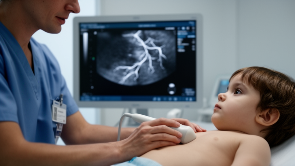
An artificial intelligence system trained to read pediatric heart ultrasounds can now find and measure the coronary arteries of young children with an accuracy that rivals the most experienced physicians, and it does so in a fraction of the time. The new deep learning model, described in the journal Pediatric Research, was developed by a team of clinicians and engineers in China to tackle one of the most stubborn problems in pediatric cardiology: reliably detecting the dangerous coronary artery damage caused by Kawasaki disease, the leading cause of acquired heart disease in children in developed countries. In head-to-head comparisons across thousands of real clinical images, the AI agreed closely with expert sonographers while cutting the average measurement time from roughly 16 to 19 seconds by hand to under two seconds by machine.
The stakes of this challenge are far higher than the small size of the vessels involved might suggest. Kawasaki disease, first described by Dr. Tomisaku Kawasaki in 1967, is an acute inflammatory condition of blood vessels that strikes primarily in early childhood and has a peculiar taste for the coronary arteries, the vessels that feed the heart muscle itself. When the disease goes untreated, the inflamed artery walls can weaken and balloon into dilations or aneurysms, which in turn set the stage for blood clots, narrowing of the vessels, and even heart attack in young patients. Because these coronary complications develop mostly during the acute phase of illness, everything depends on spotting subtle enlargements early, and then tracking vessel dimensions meticulously over years of follow-up to gauge each child’s future cardiovascular risk.
Echocardiography, the ultrasound-based imaging technique at the heart of this study, is the workhorse tool for that surveillance. It is non-invasive, reproducible, fast, and capable of showing the beating heart in real time, which makes it well suited for examining children. But it comes with a catch. Children’s coronary arteries are inherently tiny, their caliber changes as they grow, and on ultrasound they often appear with low contrast and blurred boundaries. Manual measurement of such structures leans heavily on the operator’s experience, and different observers frequently arrive at noticeably different numbers for the same vessel. That variability is problematic enough in major hospitals, but the researchers behind the new work point out that in many parts of China, and by extension in many resource-limited settings worldwide, pediatric echocardiography expertise is scarce, and achieving consistent coronary measurements is a major clinical hurdle for less experienced physicians.
To build their system, the team assembled a substantial dataset from routine clinical practice. They retrospectively collected 2,314 echocardiographic examinations, encompassing 8,766 coronary artery images, from children with suspected or diagnosed Kawasaki disease who were scanned at their hospital between January 2021 and January 2024. All the exams were performed with Philips EPIQ7 or EPIQ5 ultrasound systems while the children were asleep or calm, following the standard imaging views laid out in the American Heart Association’s guidelines. From this pool, the researchers curated 3,000 high-quality images and had two physicians with more than five years of ultrasound experience annotate four key coronary branches: the right coronary artery, the left main coronary artery, the left anterior descending artery, and the left circumflex artery. A senior pediatric echocardiographer then reviewed and corrected every annotation before the dataset was split into training, validation, and test sets in a 7:2:1 ratio.
The technical backbone of the model is YOLOv8, an open-source object detection architecture known for speed and precision. Its design splits the workload across three cooperating components. The backbone, built from so-called C2f modules, extracts visual features in alternating compression and expansion stages, using convolutions to shrink the feature maps and reduce computational load, then up-sampling to restore detail and integrate what has been learned. The neck fuses feature maps from multiple scales, combining specialized modules that let the network reason about both coarse context and fine structure, an essential capability when the targets are small, low-contrast blood vessels. The head is decoupled into separate detection and classification branches, which reduces interference between locating objects and labeling them. The researchers further tuned the pipeline for pediatric ultrasound by optimizing input resolution and data augmentation, randomly flipping, rotating, scaling, and adjusting the brightness and contrast of training images. Training ran for up to 100 epochs on an NVIDIA RTX 3090 graphics card, and a Task-Aligned Assigner along with Distribution Focal Loss helped the model cope with hard-to-detect vessels and class imbalance during learning.
Turning detection into an actual diameter measurement required an additional layer of geometric engineering. After the model locates a vessel, the software first normalizes scale, converting pixel distances into physical lengths using the calibration parameters stored in each image. It then extracts the vessel’s contour, applies a convex hull algorithm to select evenly distributed reference points, and constructs an auxiliary circle centered on each point with a radius set to half the bounding box diagonal. The intersections between that circle and the vessel outline become anchors from which perpendicular lines are drawn into the vessel interior; the lengths of these perpendiculars, averaged over multiple trials, yield the vessel diameter. The pipeline is a clever marriage of modern neural detection with classical geometry, designed specifically to handle the narrow, tortuous anatomy of pediatric coronary arteries.
When the finished model was unleashed on the test set, its performance was striking. For the right coronary artery, it achieved a recall of 96.4 percent, a precision of 94.0 percent, and a mean average precision of 99.2 percent. The left main coronary artery followed closely at 93.4 percent recall, 90.1 percent precision, and 97.0 percent mean average precision, while the left anterior descending artery scored 90.1 and 85.2 percent on recall and precision with a mean average precision of 94.1 percent. The left circumflex artery proved hardest of all, with all metrics lower than for the other branches, likely reflecting its smaller diameter, deeper anatomical position, and poorer visibility on ultrasound. Across the validation set, the model’s classifications agreed with expert physicians at a Kappa statistic of 0.904, a level conventionally interpreted as almost perfect agreement.
The comparisons with human observers were particularly revealing. Expert physicians spotted the right coronary artery and left main coronary artery in 98.4 and 98.8 percent of exams respectively, while general physicians managed 92.7 and 93.1 percent, and the gap widened dramatically for the more elusive branches: experts found the left anterior descending and left circumflex arteries in 84.9 and 57.7 percent of cases, versus just 56.0 and 37.3 percent for general physicians. Age also mattered, with recognition of the right coronary and left main arteries significantly lower in infants than in toddlers and older children, plausibly because babies’ faster breathing and heart rates complicate image acquisition. For measurements, the AI’s agreement with experts was excellent across all branches, with intraclass correlation coefficients all above 0.85 and the narrowest limits of agreement in Bland-Altman analysis, especially for the right coronary and left anterior descending arteries. Agreement between general physicians and either experts or the AI was noticeably looser, with the left circumflex artery again the weakest link for everyone. On speed, the machine was in a different league entirely: less than two seconds per measurement, compared with 16.3 seconds for experts and 18.9 seconds for general physicians using manual calipers.
The authors are candid about the study’s limitations. The model was trained and tested on static images rather than dynamic video, so it misses temporal information about moving vessels; it came from a single center using a single ultrasound brand, which may limit generalizability; the mid-segment of the right coronary artery was excluded due to sparse samples; and the bounding-box-plus-geometry approach can introduce errors in irregular vessels or poor-quality images. Future work, they say, will incorporate echocardiographic video with recurrent or transformer-based temporal modeling, refine the architecture for the difficult left anterior descending and left circumflex branches, and expand to multicenter, multi-device datasets followed by prospective clinical validation. Even so, the implications are considerable. A system that brings expert-level coronary assessment to clinics where such expertise is rare could standardize the diagnosis and follow-up of Kawasaki disease, support quality control in busy imaging departments, and help ensure that the children most at risk of lifelong cardiac damage are caught in time. The researchers caution that challenging vessels like the left circumflex artery still demand expert oversight, positioning the AI not as a replacement for skilled sonographers but as a powerful, tireless assistant, one capable of measuring a child’s coronary arteries faster than a heartbeat.
Subject of Research: A deep learning model for automated identification and measurement of coronary arteries in pediatric echocardiography for Kawasaki disease assessment
Article Title: Deep learning for coronary arteries identification and measurement in pediatric echocardiography
Article References: Deep learning for coronary arteries identification and measurement in pediatric echocardiography. (n.d.). https://doi.org/10.1038/s41390-026-05472-9
Image Credits: AI Generated
DOI: 10.1038/s41390-026-05472-9
Keywords: deep learning, Kawasaki disease, pediatric echocardiography, coronary arteries, YOLOv8, artificial intelligence, medical imaging, ultrasound, coronary artery aneurysm, object detection, cardiology, clinical AI
Cite Scienmag News
Ophelia Keating. (October 15, 2026). AI Matches Expert Doctors at Measuring Children’s Coronary Arteries on Ultrasound. Scienmag. https://scienmag.com/ai-matches-expert-doctors-at-measuring-childrens-coronary-arteries-on-ultrasound/
Ophelia Keating. "AI Matches Expert Doctors at Measuring Children’s Coronary Arteries on Ultrasound." Scienmag, 15 October 2026, https://scienmag.com/ai-matches-expert-doctors-at-measuring-childrens-coronary-arteries-on-ultrasound/. Accessed 15 October 2026.
Ophelia Keating. "AI Matches Expert Doctors at Measuring Children’s Coronary Arteries on Ultrasound." Scienmag. October 15, 2026. https://scienmag.com/ai-matches-expert-doctors-at-measuring-childrens-coronary-arteries-on-ultrasound/


