


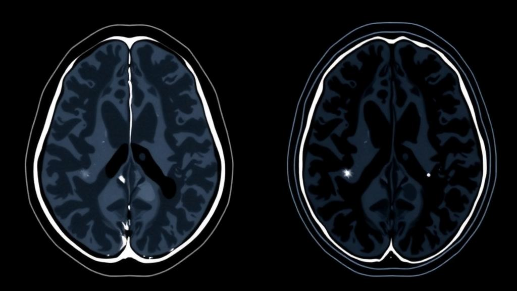
Every year, hundreds of thousands of men face the same daunting question after a prostate cancer diagnosis: how aggressive is their tumor, and how aggressive should their treatment be? The answer traditionally comes from a needle biopsy and a pathologist’s microscope, but biopsies can miss dangerous lesions and sample only a tiny fraction of the gland. A new study from researchers at Wenzhou Medical University and Zhejiang University suggests that artificial intelligence, fed with two complementary types of medical imaging, may be able to predict the aggressiveness of prostate cancer before a surgeon ever makes an incision — and, crucially, it can show its work.
The research, published in BMC Medical Imaging, tackles one of the most consequential decisions in urologic oncology: predicting the International Society of Urological Pathology, or ISUP, grade of prostate cancer before surgery. The ISUP grade, derived from the Gleason scoring system, describes how abnormal cancer cells look under a microscope and is one of the strongest predictors of how a tumor will behave. Low-grade tumors can often be monitored safely under active surveillance, while high-grade disease may demand radical prostatectomy, radiation, or systemic therapy. Getting that grade right before treatment — rather than after the gland is removed — could spare thousands of men from unnecessary surgery while ensuring that dangerous cancers are not undertreated.
The challenge is that the standard imaging tool, multiparametric magnetic resonance imaging, or mpMRI, has well-known blind spots. mpMRI excels at revealing anatomy: it shows the size and location of suspicious lesions, the integrity of the surrounding capsule, and features such as restricted water diffusion that hint at cellular density. But it offers only limited insight into tumor metabolism and molecular biology. Some biologically aggressive lesions — so-called occult high-risk disease — simply do not light up convincingly on an MRI, evading detection until pathology delivers an unwelcome surprise after surgery.
That is where the second modality comes in. The study used 18F-PSMA-1007 positron emission tomography combined with computed tomography, a molecular imaging technique that exploits a quirk of prostate cancer biology: most prostate cancer cells overexpress a protein called prostate-specific membrane antigen, or PSMA. The radiotracer 18F-PSMA-1007 binds to this protein, allowing the PET scanner to map where PSMA-hungry tumor cells are concentrated throughout the gland and the body. Because uptake intensity often correlates with tumor aggressiveness, PET/CT provides a molecular readout that MRI lacks. The trade-offs are real — higher cost and a dose of ionizing radiation — which is precisely why the researchers wanted to know whether combining the two scans with machine learning genuinely adds predictive power, rather than simply adding expense.
To answer that question, the team assembled a retrospective cohort of 341 patients who had undergone preoperative mpMRI, 18F-PSMA-1007 PET/CT, and radical prostatectomy at their institutions, along with an independent external validation cohort of 36 patients from a different center. The reference standard was uncompromising: the final ISUP grade assigned by pathologists to each surgically removed prostate. Against that gold standard, the researchers built a family of machine learning models and compared them head to head. There were five binary models — predicting simply whether a tumor was clinically significant or not — based respectively on clinical variables alone, on mpMRI alone, on PET/CT alone, on the fused imaging pair, and on the fused imaging pair plus clinical features. They also built three-class versions of the fusion models to predict full ISUP grade groups, a harder task that mirrors the granularity clinicians actually need.
The results tell a striking story about the power of multimodal fusion. In the internal cohort, a model using clinical variables alone achieved an area under the receiver operating characteristic curve, or AUC, of 0.739 — modest performance, as one would expect from variables such as age, PSA levels, and biopsy findings. The mpMRI model reached an AUC of 0.881, and the PET/CT model nearly matched it at 0.888. But when the two imaging streams were fused, performance jumped: the combined mpMRI plus PET/CT model achieved an AUC of 0.945, and adding clinical features nudged it to 0.950. In practical terms, that difference moves the model from useful to potentially transformative, because AUC values above 0.9 indicate excellent discrimination between aggressive and indolent disease — approaching the kind of reliability that could meaningfully inform treatment decisions.
The three-class task, which asks the model to distinguish low, intermediate, and high-grade disease rather than drawing a single binary line, proved predictably harder. The imaging fusion model achieved a macro-AUC of 0.810 with an accuracy of 0.695, rising to 0.830 macro-AUC and 0.698 accuracy when clinical features were added. Those numbers reflect genuine, though imperfect, discrimination across three categories — respectable for a problem where even expert pathologists sometimes disagree, but a reminder that predicting a pathology grade from pixels remains fundamentally harder than splitting tumors into two groups.
What sets this study apart from much of the medical AI literature is its insistence on interpretability. Deep learning models are notorious black boxes, and radiologists are understandably reluctant to trust a prediction they cannot interrogate. The researchers deployed two complementary explanation techniques. SHAP, which stands for SHapley Additive exPlanations, quantified how much each clinical feature contributed to each prediction, borrowing a concept from game theory to divide credit fairly among inputs. Grad-CAM, short for Gradient-weighted Class Activation Mapping, generated heatmaps overlaid on the medical images, revealing exactly which regions of the prostate the neural network looked at when making its decision. If the model flags a lesion that the radiologist can see and evaluate — rather than keying in on an artifact or a scanner-specific signature — clinicians have a concrete reason to trust it, and a concrete reason to be skeptical when the highlighted regions make no anatomical sense.
The external validation results temper the enthusiasm appropriately, and the authors are candid about it. In the 36-patient independent cohort from a different center, the fused models achieved binary AUCs of 0.726 and 0.718 — a noticeable drop from the 0.945 and 0.950 seen internally — while three-class macro-AUCs held up somewhat better at 0.778 and 0.808. This kind of performance decay across institutions is one of the most common failure modes in medical imaging AI: models can inadvertently learn center-specific scanner settings, imaging protocols, or patient populations rather than generalizable biology. The authors explicitly caution that the external validation is preliminary and that the small external cohort warrants careful interpretation. It is a refreshingly honest framing in a field where inflated claims have sometimes outrun the evidence.
Where does this leave patients and their doctors? The study, registered prospectively at the Chinese Clinical Trial Registry in October 2024, positions the fused model not as a replacement for pathology — the authors emphasize that it supplements histopathology rather than supplanting it — but as a decision-support adjunct. In a plausible clinical workflow of the future, a man with a suspicious MRI and an indeterminate biopsy might undergo PSMA PET/CT as part of his standard workup, and an interpretable fusion model would combine all available evidence into a calibrated estimate of tumor aggressiveness. That estimate could help determine whether active surveillance is genuinely safe, whether a targeted biopsy should be repeated, or whether definitive treatment should proceed without delay. For men with occult high-grade disease hiding from conventional MRI, the molecular signal from PSMA imaging, interpreted by an algorithm trained on hundreds of surgically confirmed cases, could be the difference between catching an aggressive cancer early and discovering it too late.
The road to that future runs through prospective, multicenter validation, and the authors say as much. Larger and more diverse external cohorts will be needed to confirm that the performance gains survive contact with different scanners, protocols, and populations, and the modest external results suggest that domain generalization remains the central technical hurdle. Still, the study offers a compelling proof of concept that anatomical, functional, and molecular information are genuinely complementary — that the whole of two imaging modalities, intelligently fused and transparently explained, exceeds the sum of its parts. As prostate cancer affects millions of men worldwide, an AI that can predict tumor aggressiveness before surgery, and show radiologists exactly why it reached its conclusion, represents a meaningful step toward precision medicine in one of the most common cancers on Earth.
Subject of Research: Preoperative prediction of ISUP grade in prostate cancer using interpretable multimodal fusion of 18F-PSMA-1007 PET/CT and mpMRI with machine learning
Article Title: Interpretable multimodal fusion of 18F-PSMA-1007 PET/CT and mpMRI for preoperative ISUP grade prediction in primary prostate cancer
Article References: Zhong, J., Zhou, Y., Cheng, W., Wu, T., Yuan, Y., Yao, F., Zhuang, Y., Lin, Q., Li, T., Yang, Y., Lin, Y., & Ye, Y. (2026). Interpretable multimodal fusion of 18F-PSMA-1007 PET/CT and mpMRI for preoperative ISUP grade prediction in primary prostate cancer. BMC Medical Imaging. https://doi.org/10.1186/s12880-026-02752-y
Image Credits: AI Generated
DOI: 10.1186/s12880-026-02752-y
Keywords: prostate cancer, ISUP grade, 18F-PSMA-1007 PET/CT, multiparametric MRI, multimodal fusion, machine learning, deep learning, SHAP, Grad-CAM, medical imaging, radiomics, decision support
Cite Scienmag News
Ophelia Keating. (October 10, 2026). AI Fuses PET and MRI Scans to Predict Prostate Cancer Aggressiveness Before Surgery. Scienmag. https://scienmag.com/ai-fuses-pet-and-mri-scans-to-predict-prostate-cancer-aggressiveness-before-surgery/
Ophelia Keating. "AI Fuses PET and MRI Scans to Predict Prostate Cancer Aggressiveness Before Surgery." Scienmag, 10 October 2026, https://scienmag.com/ai-fuses-pet-and-mri-scans-to-predict-prostate-cancer-aggressiveness-before-surgery/. Accessed 10 October 2026.
Ophelia Keating. "AI Fuses PET and MRI Scans to Predict Prostate Cancer Aggressiveness Before Surgery." Scienmag. October 10, 2026. https://scienmag.com/ai-fuses-pet-and-mri-scans-to-predict-prostate-cancer-aggressiveness-before-surgery/


