

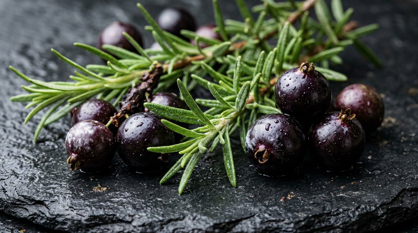
Hydroethanolic extracts derived from Salvia rosmarinus and Plinia cauliflora have shown distinct antioxidant, antibacterial, and antibiofilm properties against key microbial pathogens associated with persistent endodontic infections, according to laboratory evaluations conducted by dental researchers. The in vitro study demonstrated that these botanical preparations, prepared from rosemary leaves and jabuticaba fruit peels using a balanced hydroethanolic maceration method, exerted substantial inhibitory and bactericidal effects against planktonic cultures and mature biofilms of Enterococcus faecalis, Porphyromonas gingivalis, and Fusobacterium nucleatum. In addition to measuring microbial inhibition, the investigators characterized the specific chemical compositions of both extracts through chromatographic profiling, quantified their free radical scavenging capabilities, and assessed their biocompatibility using cultured human keratinocytes. The findings revealed that while each plant extract exhibited independent antimicrobial activity, combining specific concentrations of both botanicals yielded an additive antibacterial effect against a clinical strain of Enterococcus faecalis, providing initial foundational data on plant-derived alternatives for endodontic disinfection.
The investigation addressed an ongoing clinical obstacle in endodontology, where microbial colonization of the anatomical root canal system serves as the primary etiology of pulpal necrosis and periapical pathosis. Although conventional root canal therapy achieves clinical success rates reaching approximately ninety-seven percent, treatment failures frequently occur when microbial species survive chemomechanical disinfection and recolonize periradicular tissues to form persistent secondary infections. In these compromised root canal environments, persistent microorganisms typically organize into resilient biofilms characterized by reduced ecological diversity compared to primary dental infections. Enterococcus faecalis represents the most prevalent bacterial isolate recovered from failed endodontic procedures, largely attributable to its robust physiology and capacity to withstand adverse nutrient-depleted conditions. Furthermore, Gram-negative anaerobic pathogens including Porphyromonas gingivalis and Fusobacterium nucleatum routinely persist throughout various retreatment stages. While sodium hypochlorite remains the primary therapeutic irrigant due to broad antimicrobial efficacy and tissue-dissolving capacity, its pronounced cytotoxicity upon periapical extrusion necessitates the exploration of biocompatible alternatives.
To evaluate the therapeutic potential of botanical agents, researchers prepared hydroethanolic extracts from dried rosemary leaves and jabuticaba peels collected from local agricultural producers in São José dos Campos, Brazil. Maceration was conducted by immersing thirty grams of dried plant material into three hundred milliliters of a fifty-fifty volumetric mixture of absolute ethanol and ultrapure water obtained through a dedicated purification system. The extraction proceeded at room temperature for forty-eight hours under continuous mechanical agitation using a laboratory shaker to equilibrate polar and nonpolar bioactive recovery while preserving thermolabile secondary metabolites. Following maceration, the crude suspensions underwent sequential filtration, beginning with coarse paper filters to eliminate insoluble plant debris, followed by biological sterilization through zero-point-twenty-two micrometer membrane filtration units. The resulting sterile liquid extracts, exhibiting a dark green hue for rosemary and a deep reddish-purple coloration for jabuticaba, were preserved at four degrees Celsius prior to comprehensive analytical and biological profiling.
Phytochemical evaluation established marked quantitative and qualitative distinctions between the two hydroethanolic preparations across multiple analytical benchmarks. Soluble solids quantification yielded nine-point-forty-eight milligrams per milliliter in the Salvia rosmarinus extract and twenty-two-point-twenty-two milligrams per milliliter in the Plinia cauliflora preparation. Spectrophotometric assays utilizing standardized calibration curves determined that the rosemary extract contained one-point-fifty-eight milligrams of quercetin equivalents per milliliter of total flavonoids, whereas flavonoid concentrations in jabuticaba remained below analytical detection thresholds. Total phenolic content was measured at seven-point-forty-five milligrams of gallic acid equivalents per milliliter for rosemary and seven-point-twenty-nine milligrams for jabuticaba. High-performance liquid chromatography coupled with diode array detection at three hundred twenty nanometers identified five distinct compounds in rosemary: caffeoylquinic acid, gallotannin, chlorogenic acid, p-coumaric acid, and a quinone compound. Conversely, chromatographic fingerprinting of jabuticaba revealed four primary constituents, specifically gallic acid, p-coumaric acid, a cinnamic acid derivative, and ellagic acid, confirming the preservation of diverse bioactive polyphenol classes.
Antioxidant evaluations demonstrated that both botanical extracts effectively neutralized synthetic free radicals in laboratory spectrophotometric assays. The suppressive activity was measured by monitoring the reduction of 2,2-diphenyl-1-picrylhydrazyl in ethanol solutions across a defined concentration series, recording absorbance alterations at five hundred seventeen nanometers following hydrogen atom donation. Rosemary extract exhibited superior radical scavenging potency, generating a half-maximal effective concentration of fifty-nine micrograms per milliliter, corresponding to zero-point-zero-fifty-nine milligrams per milliliter. In comparison, jabuticaba extract required a concentration of one hundred fifty-nine micrograms per milliliter to suppress fifty percent of radical species. The robust antioxidant capacity of rosemary was primarily attributed to its high concentration of caffeoylquinic and p-coumaric acid derivatives, both known for donating hydrogen to unstable free radical intermediates.
Bactericidal profiling against planktonic bacterial suspensions revealed that both extracts suppressed viable bacterial growth across the evaluated oral strains. Broth microdilution tests could not yield reliable minimum inhibitory concentrations due to extract opacity and particle sedimentation obstructing optical turbidity readings. Consequently, researchers plated treated suspensions onto growth agar to quantify minimum bactericidal concentrations, defined as the lowest concentration preventing visible colony growth without regrowth. Rosemary extract demonstrated minimum bactericidal concentrations ranging from zero-point-fourteen to one-point-eighteen milligrams per milliliter, whereas jabuticaba bactericidal thresholds ranged between two-point-seventy-seven and five-point-fifty-five milligrams per milliliter. Checkerboard microdilution assays utilizing fractional bactericidal concentration calculations established that combining zero-point-zero-thirty-six milligrams per milliliter of rosemary with two-point-seventy-seven milligrams per milliliter of jabuticaba produced an additive antibacterial effect against the clinical strain of Enterococcus faecalis. However, no synergistic or additive interactions were detected against Porphyromonas gingivalis or Fusobacterium nucleatum, indicating strain-specific pharmacodynamic behavior.
When tested against seven-day mature biofilms of Enterococcus faecalis cultivated in microtiter plates, the botanical extracts demonstrated divergent temporal dynamics and metabolic suppression levels. Microbial metabolic activity was quantified using the colorimetric MTT reduction assay at five hundred seventy nanometers following five-minute and twenty-four-hour exposure intervals. After five minutes, application of one-point-eighteen milligrams per milliliter of rosemary extract reduced biofilm metabolic activity by fifty-two percent, while positive control treatment with two-point-five percent sodium hypochlorite achieved ninety-two percent suppression. In contrast, the additive combination of zero-point-zero-thirty-six milligrams per milliliter rosemary and two-point-seventy-seven milligrams per milliliter jabuticaba failed to produce statistically significant metabolic reductions at five minutes. Following twenty-four hours of continuous treatment, the combined extract, individual jabuticaba at five-point-fifty-five milligrams per milliliter, and rosemary at one-point-eighteen milligrams per milliliter significantly diminished biofilm viability, achieving metabolic reductions of six percent, twenty-three percent, forty percent, and eighty-eight percent across the evaluated test groups.
Antibiofilm assays against strict anaerobic oral species demonstrated distinct efficacy patterns across the evaluation intervals. For seven-day preformed Porphyromonas gingivalis biofilms, all tested concentrations of rosemary extract showed minimal efficacy at five minutes but achieved significant metabolic suppression after twenty-four hours of exposure. Conversely, all tested concentrations of jabuticaba extract reduced Porphyromonas gingivalis metabolic activity by thirty-one to forty-one percent within five minutes, demonstrating effectiveness comparable to sodium hypochlorite except at three times the bactericidal threshold. Against Fusobacterium nucleatum biofilms, all tested concentrations of both extracts significantly decreased metabolic viability after five minutes and twenty-four hours, matching the inhibitory performance of the sodium hypochlorite control. Interestingly, higher rosemary concentrations appeared to provoke increased biofilm activity in Fusobacterium nucleatum at five minutes, which the authors hypothesized might represent a hormetic defense response or inadvertent matrix stimulation triggered by sublethal phytochemical exposure, illustrating the nuanced microbial behaviors provoked by concentrated plant phenolics.
Biocompatibility assessments were conducted on spontaneously immortalized human keratinocytes cultured in Dulbecco’s Modified Eagle Medium supplemented with ten percent fetal bovine serum to evaluate epithelial cellular tolerance. Keratinocytes seeded at twenty thousand cells per well were exposed to extract dilutions for five minutes or twenty-four hours, followed by mitochondrial activity quantification via the MTT colorimetric assay. At concentrations corresponding to their respective minimum bactericidal levels and higher, both rosemary and jabuticaba extracts significantly reduced keratinocyte metabolic activity across both exposure intervals. However, when jabuticaba extract was administered at a sub-bactericidal concentration representing one-quarter of its bactericidal baseline, cell viability was preserved without statistically significant metabolic reductions. Although the extracts exhibited cytotoxic profiles comparable to sodium hypochlorite at bactericidal strengths, the preservation of cellular metabolic activity at lower concentrations suggests a window of cellular tolerance that distinguishes these natural polyphenolic preparations from aggressive synthetic chemical irrigants used in clinical endodontics.
The investigators identified several key experimental limitations that temper the clinical interpretation of these preliminary in vitro findings. Testing botanical extracts within synthetic polystyrene microplates does not replicate the structural complexity of human root dentin, the buffering capacity of organic debris, or the architecture of mature multispecies intracanal biofilms. Methodological constraints also included the reliance on metabolic dye reduction rather than direct colony-forming unit quantification, the absence of extract-specific optical blanks, and the omission of reference antioxidant standards such as Trolox or ascorbic acid. Additionally, the potential development of microbial tolerance after repeated exposure and the exact cellular mechanisms of action remain uncharacterized. Consequently, the authors emphasized that while rosemary and jabuticaba extracts demonstrate promising antioxidant, antibacterial, and antibiofilm properties, extensive further evaluations using anatomically representative root canal models, infected dentinal tubules, and expanded tissue biocompatibility assays are necessary before clinical protocols can be safely formulated for patients.
Subject of Research: Antimicrobial and antibiofilm actions of rosemary and jabuticaba extracts against endodontic pathogens
Article Title: Chemical composition, antioxidant and antibiofilm actions of Salvia rosmarinus and Plinia cauliflora extracts against endodontic pathogens and cytotoxicity on human Keratinocytes
Article References: Diamantino, M. G. G., de Carvalho, L. S., de Menezes, R. T., Rocha, C. T., Diamantino, P. S., Marcucci, M. C., Valera, M. C., Karkoutly, M., Abu Hasna, A., de Oliveira, L. D., & Carvalho, C. A. T. (2026). Chemical composition, antioxidant and antibiofilm actions of Salvia rosmarinus and Plinia cauliflora extracts against endodontic pathogens and cytotoxicity on human Keratinocytes. International Microbiology. https://doi.org/10.1007/s10123-026-00919-9
Image Credits: AI Generated
DOI: 10.1007/s10123-026-00919-9
Keywords: Endodontics, Salvia rosmarinus, Plinia cauliflora, Biofilms, Enterococcus faecalis, Antimicrobial activity, Cytotoxicity, Phytotherapy, Chemical, composition, antioxidant, antibiofilm
Cite Scienmag News
Drew Townsend. (November 7, 2026). Rosemary and Jabuticaba Extracts Inhibit Endodontic Pathogens in Laboratory Study. Scienmag. https://scienmag.com/rosemary-and-jabuticaba-extracts-inhibit-endodontic-pathogens-in-laboratory-study/
Drew Townsend. "Rosemary and Jabuticaba Extracts Inhibit Endodontic Pathogens in Laboratory Study." Scienmag, 7 November 2026, https://scienmag.com/rosemary-and-jabuticaba-extracts-inhibit-endodontic-pathogens-in-laboratory-study/. Accessed 8 November 2026.
Drew Townsend. "Rosemary and Jabuticaba Extracts Inhibit Endodontic Pathogens in Laboratory Study." Scienmag. November 7, 2026. https://scienmag.com/rosemary-and-jabuticaba-extracts-inhibit-endodontic-pathogens-in-laboratory-study/