


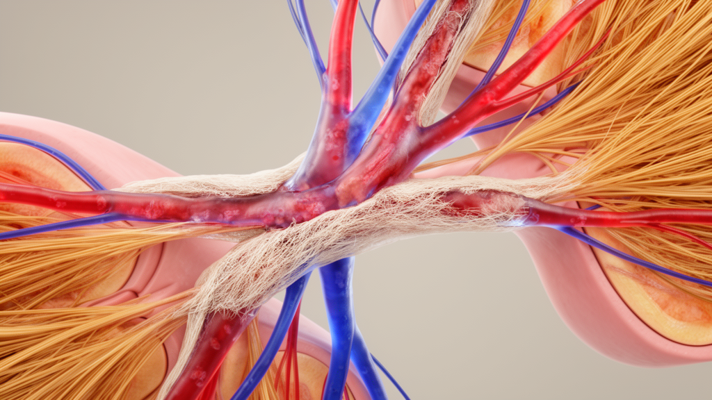
Fibrosis—excessive scarring driven by overactive fibroblasts—remains a leading cause of mortality across many diseases, yet treatment options are limited. A new review published in the Journal of Translational Medicine proposes that the missing piece of the puzzle may not lie in the scar-forming cells themselves, but in the blood vessels that supply them. The authors argue that pathological vessels act as context-dependent orchestrators of fibrotic progression, linking vascular dysfunction directly to fibrogenesis in organs throughout the body.
The review, led by Yiheng Ju and Ruoqing Xu with senior author Tao Zan, builds its framework around a central idea: aberrant vessels arise when endothelial cells fail to regain their normal quiescent state under chronic stress and instead slip into a dysfunctional mode. In healthy tissue, endothelial cells lining the vasculature alternate between active growth and rest. Under persistent inflammatory pressure, the authors contend, this balance breaks down, and the resulting dysfunctional vessels actively shape the fibrotic environment around them.
From the accumulated evidence, the authors identify recurring patterns of vascular remodeling that appear across fibrotic diseases. These include capillary de-specialization, in which highly specialized capillaries lose their organ-specific features; abortive angiogenesis, where new vessel growth is initiated but never properly completed; and venous encroachment, in which venous structures intrude on surrounding tissue architecture. Each of these remodeling modes impairs oxygen delivery, creating a hypoxic microenvironment. That oxygen-starved setting then remodels cellular behavior and fuels fibrogenesis, providing a mechanistic bridge between failing vessels and accumulating scar tissue.
Beyond oxygen deprivation, the review highlights a second channel of influence: angiocrine signaling, the chemical messages that vessels send to neighboring cells. When this signaling is disrupted, the authors argue, it can subvert the cellular communication network within the vascular niche, shifting the local environment toward a pro-fibrotic state. In this view, vessels are not passive plumbing but active participants in determining whether injured tissue resolves or scars.
To explain why fibrosis tends to persist and worsen over time, the authors integrate systemic concepts of vascular aging, proposing a persistent mismatch between vascular function and tissue demand. As vessels age, their capacity to support tissue needs may fall further out of step with what those tissues require, sustaining the chronic stress conditions under which endothelial cells remain dysfunctional. This framing connects organ-level fibrotic disease to a whole-body vascular process.
The practical implication the authors draw is a shift in therapeutic strategy: rather than targeting fibroblasts alone, next-generation anti-fibrotic therapies should aim at what they call precision vascular refunctionalization—restoring dysfunctional vessels to a healthier state in a context-specific manner. They present this as a roadmap for future drug development rather than a validated treatment approach.
It is important to note the nature of the evidence. This is a review article, published as an open-access peer-reviewed accepted manuscript, that synthesizes and interprets existing research rather than presenting new experimental data. The authors themselves frame the orchestrator role of pathological vessels as a hypothesis supported by emerging evidence, and they acknowledge that the precise mechanisms linking vascular dysfunction to fibrogenesis remain fragmented. The vascular remodeling modes and the refunctionalization concept are proposed frameworks intended to guide future research, not established clinical findings. Whether refunctionalizing vessels can actually halt or reverse fibrosis in patients will require direct experimental and clinical testing that this review does not provide.
Even so, the synthesis offers a unifying lens for a field that has largely focused on fibroblasts and inflammatory signaling. By centering the vasculature, the review suggests that the same vascular failures—failed quiescence, de-specialized capillaries, incomplete angiogenesis, venous encroachment, disrupted angiocrine signals, and aging-related functional decline—may underlie scarring in many different organs, opening a common therapeutic target where none was previously emphasized.
Subject of Research: The role of vascular dysfunction and endothelial cell states in driving fibrotic progression across multiple organs
Article Title: Vascular dysfunction orchestrates fibrotic progression in multiple organs: from mechanism decoding to therapeutic roadmap
Article References: Ju, Y., Xu, R., Li, M., Mao, J., Huang, X., Li, Q., Xia, W., & Zan, T. (2026). Vascular dysfunction orchestrates fibrotic progression in multiple organs: from mechanism decoding to therapeutic roadmap. Journal of Translational Medicine. https://doi.org/10.1186/s12967-026-08983-5
Image Credits: AI Generated
DOI: 10.1186/s12967-026-08983-5
Keywords: fibrosis, vascular dysfunction, endothelial cells, angiogenesis, hypoxia, angiocrine signaling, vascular aging, capillary de-specialization, anti-fibrotic therapies, fibroblast activation, review article, Vascular
Cite Scienmag News
Ophelia Keating. (October 27, 2026). Diseased Blood Vessels May Drive Fibrosis Across Multiple Organs. Scienmag. https://scienmag.com/diseased-blood-vessels-may-drive-fibrosis-across-multiple-organs/
Ophelia Keating. "Diseased Blood Vessels May Drive Fibrosis Across Multiple Organs." Scienmag, 27 October 2026, https://scienmag.com/diseased-blood-vessels-may-drive-fibrosis-across-multiple-organs/. Accessed 27 October 2026.
Ophelia Keating. "Diseased Blood Vessels May Drive Fibrosis Across Multiple Organs." Scienmag. October 27, 2026. https://scienmag.com/diseased-blood-vessels-may-drive-fibrosis-across-multiple-organs/


