


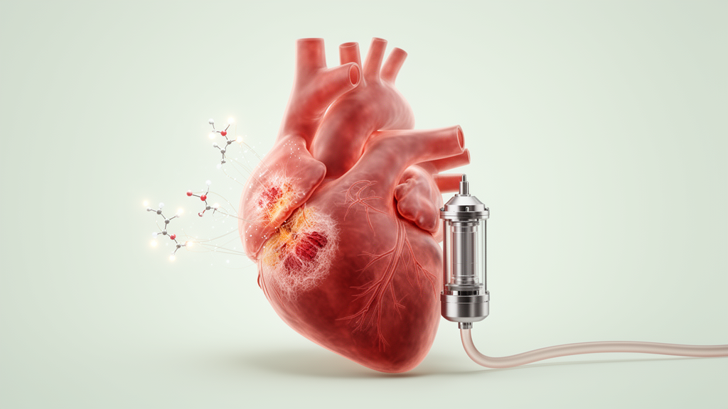
When a heart attack scars the left ventricle, the muscle that survives often begins a slow, destructive remodeling process that can end in end-stage heart failure. For a small number of these patients, a surgically implanted mechanical pump known as a left ventricular assist device, or LVAD, takes over much of the work of circulation, either as a bridge to transplantation or as long-term destination therapy. A new open-access study published in Molecular Biology Reports asks a deceptively simple question about that therapy: does unloading the failing heart with an LVAD reverse the molecular fingerprint of ischemic cardiomyopathy, or do the underlying gene and microRNA changes persist? The answer, based on paired tissue samples from eleven patients, is that many of the transcriptional abnormalities remain, and the epigenetic signals linked to cell death and scarring appear to sustain them.
The research team, led by Kajari Dhar and colleagues at the University of Nebraska Medical Center, analyzed heart muscle collected from the same patients at two time points: once when the LVAD was implanted and again when the patients ultimately underwent heart transplantation. They compared these paired pre- and post-LVAD samples with tissue from six non-failing donor hearts obtained from people who had died of brain death but had normal cardiac function on imaging. The study population was 90 percent male and 90 percent Caucasian, with an average age of 60.5 years, an ejection fraction of roughly 19 percent at the time of LVAD placement, and a mean duration of mechanical support of about 355 days. Most patients carried additional diagnoses common in advanced heart failure, including chronic kidney disease, type 2 diabetes, atrial fibrillation, and hypertension.
To profile gene activity, the investigators used the NanoString nCounter system, a hybridization-based platform that counts individual RNA molecules directly using color-coded barcoded probes, without the amplification steps required by conventional sequencing. After filtering, 127 probes from a targeted heart failure gene panel and 595 of 800 microRNA probes remained for analysis. Differential expression was assessed with the Limma statistical framework, and p-values were corrected for multiple testing using the Benjamini-Hochberg false discovery rate method. For messenger RNA, genes with a p-value below 0.05 and a fold change of at least 1.5 were called significant, while microRNAs required an adjusted p-value below 0.05 and at least a two-fold change. The team then ran Ingenuity Pathway Analysis, a computational tool that maps differentially expressed molecules onto known biological signaling networks to identify which upstream pathways are activated or suppressed.
The messenger RNA results were striking in their persistence. Compared with non-failing controls, 33 genes were differentially expressed in pre-LVAD tissue and 32 in post-LVAD tissue, and the same key genes appeared in both comparisons. The most substantially downregulated genes included BMAL1, a core circadian clock component; MYH6, which encodes the alpha myosin heavy chain central to contractile function; KCNIP2, a potassium channel modulator; and ATP2A2, the sarco/endoplasmic reticulum calcium ATPase that drives cardiac relaxation. The most significantly upregulated genes were KCND2, a potassium voltage-gated channel subunit; CRY2, another circadian regulator; and ERCC4, a DNA repair gene. Critically, none of these changes improved after LVAD implantation, meaning the mechanical unloading of the ventricle did not restore the expression of genes essential for electrical signaling, calcium handling, and contraction.
Pathway analysis added context to these persistent changes. In pre-LVAD tissue relative to non-failing controls, the signals of transforming growth factor beta 1, with a z-score of 2.65, and tumor necrosis factor alpha, with a z-score of 2.16, were both activated, along with a blood pressure disorder pathway. TGF-beta1 governs cell growth, motility, and programmed cell death, while TNF-alpha is a cytokine central to inflammatory responses, and both have been implicated in post-infarction remodeling. In post-LVAD tissue, the norepinephrine signaling and cell migration pathways were activated relative to controls. However, when the researchers compared post-LVAD directly with pre-LVAD samples from the same patients, they observed some genuinely favorable shifts: inflammatory response signaling was inhibited, and apoptosis of cardiomyocytes was inhibited with a p-value of 0.000146. Interleukin-1 beta, which was elevated before LVAD implantation, was significantly downregulated afterward, and the authors attribute this inhibition partly to increased expression of STAT3 and nitric oxide synthase alongside the drop in IL-1 beta.
A handful of genes did change meaningfully with mechanical support. Nine genes were upregulated and one downregulated in post-LVAD compared with pre-LVAD tissue. Among the upregulated genes, PDK4, a key regulator of glycolysis, and PER1, a circadian gene involved in metabolism and the cell cycle whose overexpression promotes apoptosis, showed the strongest increases, with p-values of 0.005 and 0.006 respectively. The authors note that PDK4 and PER1 elevation with LVAD therapy mirrors their earlier findings in non-ischemic cardiomyopathy, suggesting a shared metabolic and circadian response to unloading across heart failure etiologies.
The microRNA data formed the epigenetic heart of the study. Relative to non-failing controls, 195 microRNAs were differentially expressed in pre-LVAD tissue and 120 in post-LVAD tissue, yet a scatter plot comparison of fold changes across all 800 measured microRNAs revealed a correlation of 0.922 between the two states, indicating that the global microRNA landscape was nearly identical before and after mechanical unloading. Ingenuity Pathway Analysis predicted that upregulated microRNAs activated apoptosis in both pre-LVAD tissue, based on 27 differentially expressed microRNAs with a z-score of 0.766, and post-LVAD tissue, based on 15 microRNAs with a z-score of 2.155, meaning the pro-apoptotic signal actually intensified with support. The same analysis predicted that eight microRNAs, including miR-16-5p, miR-30b-5p, miR-23a-3p, miR-208b-3p, miR-125a-5p, miR-29b-3p, miR-21-5p, and miR-150-5p, were associated with cardiac fibrosis in both pre- and post-LVAD states, a prediction the team corroborated by visualizing fibrosis in patient tissue with Masson’s trichrome staining.
Several of these microRNAs carry particular weight in cardiac biology. MiR-208b belongs to a heart-enriched family encoded within the myosin heavy chain genes, and members of this family regulate the myosin isoform switching that occurs during development and in diseased hearts. MiR-23a, miR-29, and miR-30 family members have been linked to arrhythmia, dilated and hypertrophic cardiomyopathy, and myocardial infarction in prior work. To connect the two RNA layers, the researchers performed an integrated analysis restricted to inverse relationships, where an upregulated microRNA paired with a downregulated messenger RNA or vice versa. In pre-LVAD tissue, 124 microRNAs were predicted to alter the expression of 24 genes, while in post-LVAD tissue 12 microRNAs were linked to 12 genes, with several microRNAs targeting more than one transcript. This regulatory web offers a mechanistic explanation for why messenger RNA changes outlast the period of ventricular unloading.
To guard against platform-specific artifacts, the team validated their targeted NanoString results using unbiased messenger RNA sequencing on the AVITI24 system from Element Biosciences, sequencing 23 samples comprising four non-failing controls, nine pre-LVAD, and ten post-LVAD tissues. All samples passed quality thresholds for uniquely mapped reads against the human GRCh38 reference genome, and differential expression was computed with DESeq2 using a negative-binomial generalized linear model. The sequencing run confirmed the downregulation of MYH6, ATP2A2, BMAL1, and KCNIP2 and the upregulation of KCND2, ERCC4, and CRY2 in both pre- and post-LVAD tissue, as well as the post-LVAD increases in PDK4 and PER1, yielding highly concordant results across the two technologies.
The authors are careful to frame the scope of what this study can and cannot show. With only eleven paired patients, mostly male and Caucasian, and with pre-LVAD samples taken from the left ventricular apex while post-LVAD and control samples came from the ventricular free wall, sampling geometry and demographic narrowness both limit generalizability. As a retrospective observational study, changes in medical therapy over the months of LVAD support could also have influenced gene expression, and the targeted panel by design captured only heart failure-associated transcripts. Limited tissue availability prevented direct measurement of apoptosis by cleaved caspase-3 Western blotting or TUNEL staining. Even so, the central conclusion stands on the paired data: in end-stage ischemic cardiomyopathy, microRNA expression is tied to cell death and fibrosis pathways, those abnormal patterns largely remain after LVAD support, and they may help sustain the messenger RNA changes that keep the unloaded heart in a state of ongoing failure. For a therapy that rescues survival, the molecular record of the infarcted heart appears remarkably resistant to erasure.
Subject of Research: mRNA and microRNA expression profiles in ischemic cardiomyopathy before and after LVAD support
Article Title: Analysis of mRNA and microRNA profiles in ischemic cardiomyopathy with LVAD support
Article References: Dhar, K., Basma, H., Qiu, F., Um, J. Y., Anderson, D. R., & Lowes, B. D. (2026). Analysis of mRNA and microRNA profiles in ischemic cardiomyopathy with LVAD support. Molecular Biology Reports, 53(1), Article 1816. https://doi.org/10.1007/s11033-026-13007-y
Image Credits: AI Generated
DOI: 10.1007/s11033-026-13007-y
Keywords: ischemic cardiomyopathy, LVAD, microRNA, mRNA expression, cardiac remodeling, heart failure, apoptosis, fibrosis, NanoString, RNA sequencing, Ingenuity pathway analysis, translational research
Cite Scienmag News
Juliet Wilcox. (October 26, 2026). MicroRNA Scars of Heart Failure Persist After LVAD Support in Ischemic Cardiomyopathy. Scienmag. https://scienmag.com/microrna-scars-of-heart-failure-persist-after-lvad-support-in-ischemic-cardiomyopathy/
Juliet Wilcox. "MicroRNA Scars of Heart Failure Persist After LVAD Support in Ischemic Cardiomyopathy." Scienmag, 26 October 2026, https://scienmag.com/microrna-scars-of-heart-failure-persist-after-lvad-support-in-ischemic-cardiomyopathy/. Accessed 26 October 2026.
Juliet Wilcox. "MicroRNA Scars of Heart Failure Persist After LVAD Support in Ischemic Cardiomyopathy." Scienmag. October 26, 2026. https://scienmag.com/microrna-scars-of-heart-failure-persist-after-lvad-support-in-ischemic-cardiomyopathy/


