


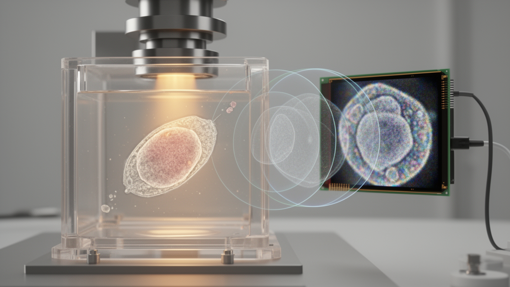
A deep-learning reconstruction network called XPAR-Net improved average reconstruction quality over HoloFormer on a complete 11-category in-distribution test set for lens-free on-chip microscopy. For a 256 × 256 input patch, XPAR-Net increased average peak signal-to-noise ratio by 1.28 dB and average structural similarity by 2.9% relative to HoloFormer. The comparison also found that XPAR-Net used only 8.8% of HoloFormer’s trainable parameters and 19.0% of its multiply-accumulate operations under the evaluated conditions.
Lens-free on-chip microscopy is a compact, wide-field imaging method, but reconstructing a complex optical field from a single in-line hologram presents two coupled challenges. The measurement records intensity rather than the complex field directly, requiring the reconstruction system to infer phase. It must also recover fringe-scale information within the finite numerical-aperture bandwidth of the acquisition system. XPAR-Net was designed to address these tasks by coordinating learning across complementary representation paradigms rather than relying on a single type of feature representation.
The network combines field-wide channel-relation modeling with localized spatial refinement. The first component captures selective dependencies among holographic responses across the field, while the second preserves fine structural variations. A Cross-Domain Alignment and Fusion module reconciles the two representations. XPAR-Net also confines learnable spectral residual correction to the modeled acquisition support, an architectural constraint that limits where the network can apply such correction. Training couples reference-domain complex-field fidelity with in-support spectral-profile, local-structural and input-relative refinement constraints.
These results support the evaluated model’s combination of reconstruction accuracy and computational compactness: XPAR-Net outperformed HoloFormer on the reported test set while requiring a small fraction of its trainable parameters and multiply-accumulate operations for a 256 × 256 input patch. The evidence applies to the study’s 11-category in-distribution evaluation and compared conditions; the supplied report does not establish performance for other test distributions. The article was published on 21 October 2026 in Scientific Reports as an early, peer-reviewed version that remains subject to further editing before the final version of record replaces it.
Subject of Research: Evaluation of XPAR-Net for single-frame lens-free on-chip microscopy reconstruction
Article Title: XPAR-Net: cross-paradigm alignment and reconstruction network for high-fidelity lens-free on-chip microscopy
Article References: Zhao, Y., Su, X., Xu, N., & Gao, Y. (2026). XPAR-Net: cross-paradigm alignment and reconstruction network for high-fidelity lens-free on-chip microscopy. Scientific Reports. https://doi.org/10.1038/s41598-026-73646-w
Image Credits: AI Generated
DOI: 10.1038/s41598-026-73646-w
Keywords: XPAR-Net, lens-free microscopy, on-chip imaging, holographic reconstruction, deep learning, computational imaging, phase retrieval, optical fields, image reconstruction, neural networks, cross-paradigm, alignment
Cite Scienmag News
Blake Davidson. (October 26, 2026). XPAR-Net: cross-paradigm alignment and reconstruction network for high-fidelity lens-free on-chip microscopy. Scienmag. https://scienmag.com/xpar-net-cross-paradigm-alignment-and-reconstruction-network-for-high-fidelity-lens-free-on-chip-microscopy/
Blake Davidson. "XPAR-Net: cross-paradigm alignment and reconstruction network for high-fidelity lens-free on-chip microscopy." Scienmag, 26 October 2026, https://scienmag.com/xpar-net-cross-paradigm-alignment-and-reconstruction-network-for-high-fidelity-lens-free-on-chip-microscopy/. Accessed 26 October 2026.
Blake Davidson. "XPAR-Net: cross-paradigm alignment and reconstruction network for high-fidelity lens-free on-chip microscopy." Scienmag. October 26, 2026. https://scienmag.com/xpar-net-cross-paradigm-alignment-and-reconstruction-network-for-high-fidelity-lens-free-on-chip-microscopy/


