


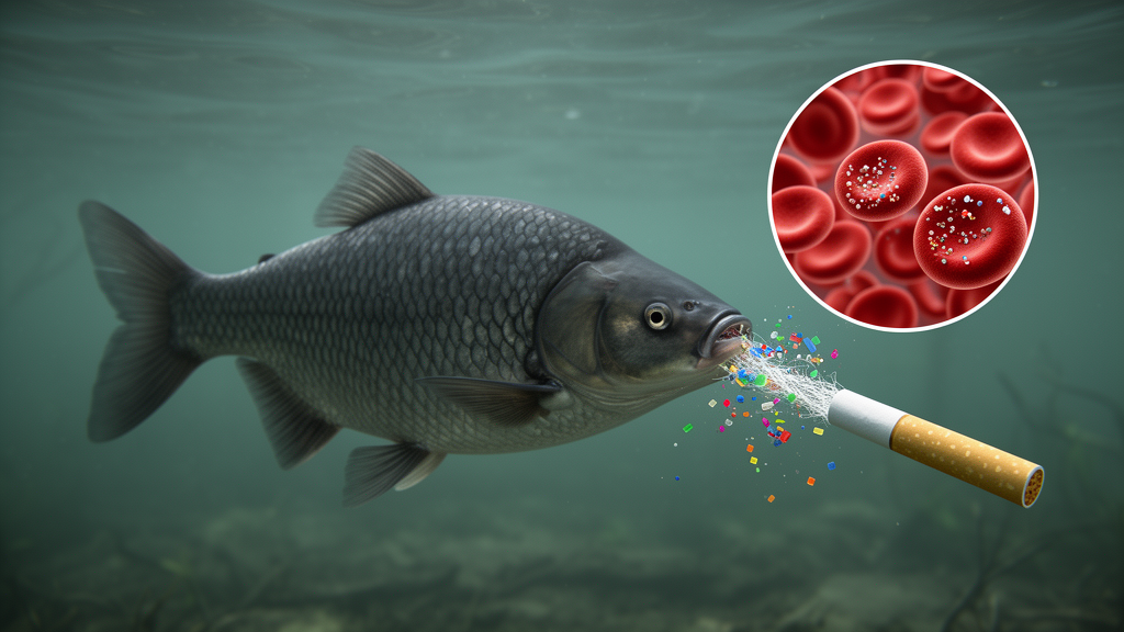
Cigarette filters, one of the world’s most ubiquitous pieces of plastic litter, have just been shown to do something unexpected when they are broken down to the nanoscale. In a laboratory experiment in Brazil, juvenile tambaqui — the Amazon’s iconic Colossoma macropomum — exposed to cellulose acetate nanoparticles for as little as one hour developed measurable shifts in their blood chemistry, from falling hemoglobin and serum proteins to rising glucose and urea. The fish survived every concentration tested, and many of the changes faded after the animals were moved to clean water. But the speed of the response, and the fact that some of it persisted even after the particles were removed, is drawing attention to a polymer that has long flown under the radar of nanoplastic research.
The study, published in the journal Ecotoxicology, focused on a material with an enormous environmental footprint. Cellulose acetate is one of the most commercially important cellulose-derived polymers, used in textiles, biomedical films and, above all, cigarette filters. Global plastic production has exploded from roughly 2 million tons in 1950 to an estimated 400.3 million tons in 2022, and freshwater ecosystems act as receptors for pollutants draining from entire river basins. Cellulose acetate is among the most common plastic contaminants found in aquatic environments, especially marine ones. It is considered resistant to biodegradation, yet it is photodegradable: under ultraviolet radiation it fragments into micro- and nanoplastics that can persist in the aquatic environment for up to 15 years. Cellulose acetate nanoparticles are also used as carriers of agricultural inputs and can flow directly into water bodies.
The research team chose tambaqui deliberately. The species, a member of the family Characidae, is native to the Amazon basin, where previous work has documented microplastics in the stomach contents of fish spanning herbivore, omnivore and carnivore trophic guilds. Plastic particles are classified by size as macroplastics larger than 5 millimeters, microplastics between 0.1 and 5 millimeters, and nanoplastics smaller than 1000 nanometers by some authors and smaller than 100 nanometers by others. Fish encounter these particles through gill respiration and gastrointestinal ingestion, and nanoparticles can cross epithelia by endocytosis to reach the circulatory system. Once in circulation, they can accumulate in vital organs such as the intestine, liver and brain because they are able to cross cell membranes, triggering metabolic disorders, altered lipid metabolism, inflammatory responses, oxidative imbalances, liver dysfunction and reproductive interference. Blood parameters therefore serve as integrated biomarkers of these systemic effects, and particles can even interact with erythrocytes, albumin and globulins in ways that contribute to vascular obstruction.
The experimental design was tightly controlled. A total of 120 juvenile tambaqui, averaging 38.91 ± 5.57 grams and 13.76 ± 0.74 centimeters in total length, were randomly distributed into 12 tanks of 150 liters within a recirculating aquaculture system, at a density of 10 fish per tank. After 15 days of acclimatization on a commercial diet, the juveniles were exposed to one of four concentrations of cellulose acetate nanoparticles: 0 milligrams per liter as a control, 0.01, 0.10 and 1.00 milligrams per liter. The lowest concentration was chosen to encompass environmentally relevant values, while the higher levels simulated future scenarios of intensified plastic pollution. Blood was drawn after 1 hour and after 24 hours of exposure, and the fish were then relocated to a tank of clean water for another 24 hours before a final sample was collected. Each tank was designated for only one sampling time, with the tank treated as the experimental unit to avoid the physiological interference of repeated handling stress.
The nanoparticles themselves were extracted from unused cigarette filters of three commercial brands, dissolved in dichloromethane and emulsified with polyvinyl alcohol using mechanical mixing and ultrasonication. Fourier transform infrared spectroscopy confirmed cellulose acetate as the primary constituent, with characteristic absorption bands at 1737, 1371, 1219 and 1034 centimeters to the minus one, corresponding to carbonyl stretching and C–O–C vibrations. Dynamic light scattering measured an average hydrodynamic diameter of 230.86 ± 3.83 nanometers with a polydispersity index of just 0.08, indicating a remarkably uniform suspension, and a zeta potential of −21.54 millivolts, reflecting moderate colloidal stability. Atomic force microscopy revealed a bimodal size distribution, with individual particles averaging 181 ± 13 nanometers and agglomerates averaging 435 ± 24 nanometers. The authors note that the difference between dry-particle and hydrodynamic measurements is expected, and that the sizes fall within the range capable of achieving cellular penetration in juvenile fish.
The hematological findings were strikingly rapid. After just one hour of exposure, hemoglobin was highest in the control group and lowest in fish exposed to 1.00 milligrams per liter of nanoparticles, and mean corpuscular hemoglobin concentration was similarly reduced at the highest dose. Yet by 24 hours, and again after the recovery period, these parameters showed no significant differences among treatments. Hematocrit, erythrocyte counts, mean corpuscular volume and mean corpuscular hemoglobin remained statistically unchanged throughout. No mortality was observed at any concentration during the entire experiment. Comparable reductions in hemoglobin have been reported in Nile tilapia exposed to polystyrene nanoparticles and in other species exposed to polyethylene microplastics, but this study is among the first to isolate the effects of cellulose acetate particles at the nanoscale, a gap the authors attribute to the difficulty of disentangling cigarette-butt-derived cellulose acetate from the other smoke contaminants that leachates carry.
The protein profile told a more persistent story. Serum protein and globulin levels dropped after one hour at 1.00 milligrams per liter, and the reduction in serum protein was still evident after 24 hours of exposure. Serum proteins play a crucial role in maintaining osmotic balance by preventing excessive fluid outflow from the circulation, and stress responses release adrenaline, which can increase gill permeability and alter blood plasma volume. The authors suggest that continuous exposure to the nanoparticles may have altered protein metabolism, reducing serum proteins or increasing their utilization. The decline in globulins may relate to changes in immune response described in the literature, although the study did not evaluate specific immunological biomarkers that would confirm this mechanism. Albumin was also lower at the highest concentration after one hour, with partial normalization over time.
Metabolic markers revealed a delayed stress response. Glucose did not differ among treatments after one hour, but after 24 hours the fish exposed to 0.10 and 1.00 milligrams per liter showed elevated glucose, a widely recognized indirect stress biomarker in fish. Toxic exposure mobilizes energy reserves through catecholamines and corticosteroids that stimulate glycogenolysis, and can also reduce glucose uptake while activating gluconeogenesis. Even after transfer to clean water, juveniles exposed to the highest concentration were unable to bring glucose back down, which the authors interpret as a prolonged stress response. Urea, a nitrogenous byproduct of protein metabolism, rose within the first hour at 0.10 and 1.00 milligrams per liter, consistent with stress-intensified proteolysis, but returned to normal after the recovery period. Triglycerides and cholesterol fell at 1.00 milligrams per liter after both one and 24 hours of exposure, echoing findings with polystyrene nanoparticles in tilapia, and both recovered to baseline in clean water. The activities of the transaminases AST and ALT, enzymes released when hepatocyte membranes become more permeable and important in gluconeogenesis, were altered after 24 hours; ALT normalized during recovery, but AST remained elevated at higher concentrations, indicating the enzymatic response was not completely reversed within the evaluated interval.
One of the most intriguing patterns was what the data did not show: a clear progressive relationship between increasing concentration and effect intensity. In several cases the intermediate concentration of 0.10 milligrams per liter triggered responses similar to or even more pronounced than the highest dose, a non-monotonic pattern previously described in ecotoxicological studies of plastic contaminants. The authors also flag an important limitation. Polyvinyl alcohol was used as a surfactant during synthesis and may have remained in the final suspension; with no quantification of residual PVA and no PVA-matched control group, the theoretical maximum PVA concentrations would have been 0.02, 0.20 and 2.00 milligrams per liter in the three nanoparticle treatments. Published work suggests PVA is largely benign below 1 milligram per liter, but adverse effects have been observed in zebrafish at 5 milligrams per liter, so a contribution of the surfactant, especially in the highest treatment, cannot be completely ruled out. Exposure concentrations were nominal, as analytical verification in the culture water was not technically feasible because dissolved organic matter and suspended particles interfere with nanoparticle detection.
For now, the message is one of acute, partially reversible disturbance rather than lasting damage. The absence of mortality, the normalization of urea, triglycerides, cholesterol and several hematological parameters after 24 hours in clean water, and the predominantly transient nature of the changes suggest the fish mounted compensatory physiological adjustments. But the authors caution that the responses observed after one and 24 hours cannot be extrapolated to longer exposure periods, during which nanoparticles may gradually accumulate in tissues and reach the liver, the central organ for processing potentially toxic substances. Environmental concentrations of nanoplastics remain deeply uncertain — estimates for high environmental nanoparticle levels reach about 0.015 milligrams per liter, while the experimental gradient spanned an estimated environmental scenario, a possible future scenario and an extreme scenario. Future work, the team writes, should integrate tissue-level analyses, oxidative stress parameters and histopathological assessments, increase the number of replicates, and include surfactant-matched controls to fully separate the effects of the particles from those of their synthesis vehicle.
Subject of Research: Cellulose acetate nanoparticles: impacts of concentration and exposure time on the biochemistry and hematology of juvenile Colossoma macropomum
Article Title: Cellulose acetate nanoparticles: impacts of concentration and exposure time on the biochemistry and hematology of juvenile Colossoma macropomum
Article References: de Oliveira, C. G., Boaventura, T. P., dos Santos, F. A. C., de Sena Souza, A., dos Santos Silva, S., Carvalho Ananias, I. D. M., Gil, C. S. B., Luz, R. K., & Oréfice, R. L. (2026). Cellulose acetate nanoparticles: impacts of concentration and exposure time on the biochemistry and hematology of juvenile Colossoma macropomum. Ecotoxicology, 35(9), Article 189. https://doi.org/10.1007/s10646-026-03174-w
Image Credits: AI Generated
DOI: 10.1007/s10646-026-03174-w
Keywords: Cellulose, acetate, nanoparticles, impacts, concentration, exposure, time, biochemistry, hematology, juvenile, Colossoma, macropomum
Cite Scienmag News
Bethany Barker. (October 24, 2026). Cigarette Filter Nanoplastics Rapidly Alter the Blood Chemistry of Amazon’s Tambaqui Fish. Scienmag. https://scienmag.com/cigarette-filter-nanoplastics-rapidly-alter-the-blood-chemistry-of-amazons-tambaqui-fish/
Bethany Barker. "Cigarette Filter Nanoplastics Rapidly Alter the Blood Chemistry of Amazon’s Tambaqui Fish." Scienmag, 24 October 2026, https://scienmag.com/cigarette-filter-nanoplastics-rapidly-alter-the-blood-chemistry-of-amazons-tambaqui-fish/. Accessed 24 October 2026.
Bethany Barker. "Cigarette Filter Nanoplastics Rapidly Alter the Blood Chemistry of Amazon’s Tambaqui Fish." Scienmag. October 24, 2026. https://scienmag.com/cigarette-filter-nanoplastics-rapidly-alter-the-blood-chemistry-of-amazons-tambaqui-fish/


