


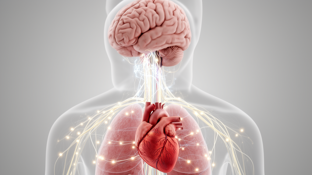
Parkinson’s disease is associated with altered coordination between brain activity and cardiac dynamics, according to a focused review of electrophysiological, neuroimaging, and physiological studies in people with the disorder. The evidence suggests that this disruption can be detected in early and untreated Parkinson’s, varies with the severity of autonomic dysfunction, and may change with dopaminergic treatment. Cross-system measures also revealed abnormalities when heart-rate variability or brain signals were considered separately. However, the review identified only seven eligible reports, representing six independent patient cohorts, and concluded that brain–heart coupling is an emerging physiological framework rather than a validated clinical biomarker.
Cardiovascular autonomic abnormalities are already well established in Parkinson’s disease. They include reduced or altered heart-rate variability, impaired baroreflex responses, unstable blood pressure, orthostatic hypotension, and loss of cardiac sympathetic innervation. These findings are not interchangeable: heart-rate variability describes modulation of cardiac rhythm, baroreflex testing evaluates a closed cardiovascular reflex, orthostatic hypotension captures a hemodynamic outcome, and cardiac sympathetic imaging assesses peripheral nerve terminals. The review argues that their partial dissociation may reflect dysfunction at several levels of the neurocardiac axis, making direct analysis of brain–heart relationships potentially more informative than any conventional measure alone.
The physiological basis is a distributed, bidirectional system. Descending signals from the central autonomic network—including the insula, cingulate cortex, amygdala, hypothalamus, brainstem, and periaqueductal gray—reach the heart through sympathetic and parasympathetic pathways. These signals interact with spinal, intrathoracic, and intrinsic cardiac neuronal networks, including the heart’s own “little brain,” which integrates local mechanical and chemical information with external autonomic input. In the other direction, cardiac mechanical, hemodynamic, and chemical signals reach the brain through vagal and spinal afferents, contributing to reflex control, arousal, emotion, cognition, and the representation of internal bodily states.
Direct evidence comes from studies that simultaneously or temporally aligned brain and cardiac measurements. In one early study, 26 people with Parkinson’s-related autonomic dysfunction and 23 controls underwent EEG and electrocardiography. Frequency-domain heart-rate variability did not clearly separate the groups, but the association between cortical oscillations and cardiac autonomic indices differed, particularly in lateral premotor–parietal circuits. Another study included 14 people with de novo, drug-naïve Parkinson’s and 14 controls. Resting-state functional MRI and dynamic high-frequency heart-rate variability showed disrupted relationships between cardiac parasympathetic fluctuations and activity in the cingulate and insular cortex, prefrontal and sensorimotor regions, supplementary motor area, hippocampus, precuneus, and brainstem.
Subsequent work reinforced the pattern. Thirty-one people with Parkinson’s and 21 controls underwent resting-state fMRI with heart-rate variability derived from photoplethysmography. A heart–brain synchronization index was reduced in Parkinson’s across regions associated with the central autonomic network, and greater desynchronization accompanied more severe dysautonomia. The measure also correlated with objective tests of sympathetic and cardiovagal function, including Valsalva pressure recovery and heart-rate responses during deep breathing. These are cross-sectional associations, however, and neither the synchronization index nor similar lagged correlations can establish the direction or directness of communication.
Simultaneous EEG–ECG recordings offered higher temporal resolution. In a study of 15 people with Parkinson’s examined both off and on dopaminergic medication and 16 healthy controls, coupling between alpha- and gamma-band connectivity and cardiac sympathetic dynamics was reduced off medication. Dopaminergic treatment partially restored the coupling, and greater recovery was associated with a larger motor improvement. Related analyses of the same overlapping dataset linked cardiac dynamics to time-varying properties of brain networks, including clustering, efficiency, modularity, and assortativity. These results show that abnormalities may reside in the relationship between systems rather than in either system alone, but medication can also alter movement, arousal, blood pressure, cortical state, and peripheral autonomic function, so a direct dopaminergic mechanism cannot be inferred.
The heart-to-brain direction remains less firmly established. Behavioral studies indicate that some people with Parkinson’s perceive their own heartbeats less accurately than healthy controls, although heartbeat-counting tasks are influenced by attention, time estimation, expectations, and knowledge of heart rate. A seven-person conference study of 64-channel EEG and ECG found altered heartbeat-evoked potentials and heart-rate variability off medication, with both moving toward control values on levodopa. Heartbeat-evoked potentials are cortical responses time-locked to individual cardiac events, but they are not direct recordings of cardiac afferent integrity. Their amplitude and topography also depend on attention, respiration, arousal, EEG referencing, artifact correction, and the selected analysis window, so the Parkinson’s findings remain hypothesis-generating.
Conventional autonomic assessment remains necessary, and brain–heart analysis is complementary rather than a replacement. Standardized cardiovascular autonomic reflex tests evaluate heart-rate response to deep breathing, the 30:15 standing ratio, the Valsalva maneuver, and blood-pressure response to standing; tilt-table testing, baroreflex analysis, heart-rate variability, and cardiac sympathetic imaging add further information. A future clinical measure would need to outperform these established tests, show test–retest reliability, work at the individual level, and clarify whether it reflects impaired descending control, altered afferent processing, or disturbed bidirectional integration. Respiration, blood pressure, posture, vigilance, movement, tremor, age, cardiovascular disease, medication, genotype, and disease stage must be measured or modeled rather than treated as irrelevant noise.
Longitudinal work is also needed because most direct studies are small and cross-sectional, and two analyses used the same Parkinson’s dataset. There is currently no evidence that brain–heart coupling can track progression or prospectively classify patients. Orthostatic stress, dopaminergic manipulation, controlled neuromodulation, and sensory reduction could help identify which links in the neurocardiac loop fail under challenge, while serial recordings could establish whether coupling abnormalities precede or follow clinically evident dysautonomia. The review concludes that Parkinson’s may disrupt not only the brain and cardiovascular system considered independently, but the dynamic relationships that integrate them. That network-physiology perspective could eventually help characterize disease heterogeneity and therapeutic response, although its diagnostic and prognostic value remains unproven.
Subject of Research: Evidence that Parkinson’s disease alters bidirectional brain–heart coupling and cardiovascular autonomic integration.
Article Title: Brain–heart coupling in Parkinson’s disease: from autonomic dysfunction to network physiology
Article References: Pagliaro, P. (2026). Brain–heart coupling in Parkinson’s disease: from autonomic dysfunction to network physiology. Frontiers in Physiology, 17, 1974885. https://doi.org/10.3389/fphys.2026.1974885
Image Credits: AI Generated
DOI: 10.3389/fphys.2026.1974885
Keywords: Parkinson’s disease, brain–heart coupling, autonomic dysfunction, cardiac interoception, heart-rate variability, network physiology, neurocardiology, dopaminergic treatment, EEG, functional MRI, Brain, heart
Cite Scienmag News
Cassandra Pierce. (October 22, 2026). Parkinson’s disrupts brain–heart coordination, review finds. Scienmag. https://scienmag.com/parkinsons-disrupts-brain-heart-coordination-review-finds/
Cassandra Pierce. "Parkinson’s disrupts brain–heart coordination, review finds." Scienmag, 22 October 2026, https://scienmag.com/parkinsons-disrupts-brain-heart-coordination-review-finds/. Accessed 22 October 2026.
Cassandra Pierce. "Parkinson’s disrupts brain–heart coordination, review finds." Scienmag. October 22, 2026. https://scienmag.com/parkinsons-disrupts-brain-heart-coordination-review-finds/


