


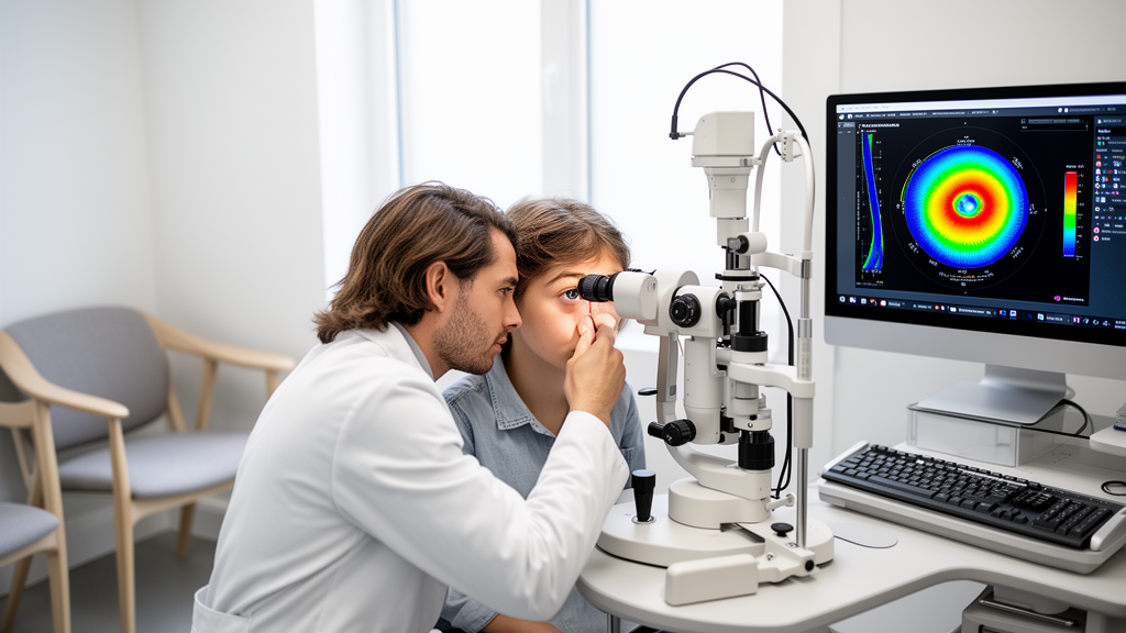
Machine learning can classify keratoconus severity and predict aspects of disease progression and surgical outcomes with promising results in retrospective studies, but a systematic review concludes that the evidence remains insufficient to establish clinical benefit. The analysis identified 41 reports published between January 2015 and October 2025: 20 addressing severity classification, 10 addressing progression assessment or prediction, and 11 addressing treatment outcomes. Much of the evidence came from retrospective, single-center studies, while independent multicenter evaluation and prospective clinical workflows remained limited.
Keratoconus is a progressive corneal ectasia disorder marked by stromal thinning, corneal protrusion and irregular myopic astigmatism. Its reported prevalence varies substantially among epidemiologic studies: one meta-analysis estimated 1.38 cases per 1,000 people, while another estimated 2.89 per 1,000, with the latter analysis reporting 5.26 per 1,000 among adults aged 20 to 29. Clinicians currently combine corneal topography and tomography, refractive measurements and visual acuity to assess severity and determine how closely patients should be monitored. Maximum keratometry, or Kmax, is commonly used to characterize corneal curvature, but its ability to identify progression depends on the threshold and reference definition chosen. Clinical staging systems such as the Amsler–Krumeich classification describe severity but do not fully capture visual function or future deterioration.
The strongest body of evidence concerned severity classification. Studies used Pentacam Scheimpflug tomography, Placido-based and combined topography systems, Corvis ST biomechanical measurements, anterior-segment optical coherence tomography and frontal or lateral corneal images. Conventional methods included logistic regression, support vector machines, random forests and discriminant analysis, while deep-learning studies employed convolutional neural networks such as ResNet, EfficientNet, Inception V3 and VGG16. One random-forest model classified 644 eyes into stages 0 to 4 with reported accuracy of 98.6%, but its comparison models were substantially less accurate on the same data. Other image-based systems reported four-class accuracies of 94.23% to 95.0%, and a model using Pentacam color-coded maps achieved an external five-class area under the receiver operating characteristic curve, or AUC, of 0.859.
These headline figures do not all measure the same kind of success. Some models reproduced labels assigned by clinicians, whereas others predicted continuous severity or clustered eyes according to device-derived measurements. Dataset units also differed, including eyes, scans, records and augmented images, making direct comparison difficult. A Pentacam clustering analysis that assigned eyes to Amsler–Krumeich stages achieved a mean accuracy of 76.9% with a standard deviation of 3% over 20 runs, and stage-specific AUCs ranged from 0.79 to 0.99. Another study reported 97.8% accuracy for classification based on frontal and lateral images, but its confusion matrix showed 250 of 268 cases correctly classified, equivalent to 93.3%; that internal discrepancy limits interpretation.
Progression research was smaller and more fragmented because the studies did not forecast the same endpoint. Some models predicted future tomographic change, others detected change that had already occurred, and several used subsequent corneal cross-linking referral or receipt as their label. A time-delay neural network given age and five tomographic indices at earlier visits predicted worsening at a third examination with reported sensitivity of 70.8% and specificity of 80.6%. A random-forest model evaluated under three Kmax-change thresholds reported AUCs of 0.90, 0.91 and 0.93 for thresholds of 0.75, 1.00 and 1.25 diopters. Because changing the threshold also changed the outcome definition, those figures cannot establish that one threshold or model was superior in predicting clinically meaningful deterioration.
Other progression studies illustrate the difference between forecasting and recognition. A paired-map convolutional network detected a progression signal a mean of 11.1 months before a conventional one-diopter change in flat or steep keratometry, although its development and evaluation cohorts consisted of 385 and 1,331 eyes, respectively, and the analysis was centered on eventual progressors. A model trained to predict subsequent cross-linking over follow-up ranging from six weeks to 8.6 years achieved a fivefold AUC of 0.814, with 77.8% sensitivity and 69.6% specificity. That endpoint reflects treatment policy as well as biological progression. In another study, serial examinations improved prediction of subsequent cross-linking referral from an AUC of 0.59 for a single examination to 0.75 for serial data, but serial precision was only 0.32 despite a recall of 0.67.
Surgical-outcome models similarly divided into distinct clinical questions. Across corneal cross-linking, intracorneal ring segment implantation and keratoplasty, targets included changes in corrected visual acuity, Kmax, corneal thickness, postoperative maps, graft status and rejection. An XGBoost model evaluated in a later cohort from the same center predicted two-year post-cross-linking changes in visual acuity and Kmax with coefficients of determination of 0.8956 and 0.8382, respectively. A LightGBM model estimated minimum and central corneal thickness with test root mean squared errors of 19.22 and 19.68 micrometers. For ring implantation, one CatBoost model reported an acuity coefficient of determination of 0.59 and mean absolute error of 1.08 diopters for mean keratometry, indicating that the task remained difficult despite favorable aggregate correlations.
Limited comparative studies suggested possible value for model-guided treatment planning, but their design prevents firm causal conclusions. In a small, nonrandomized comparison involving two groups of 20 eyes, artificial-neural-network-guided ring implantation was associated with better three-month corrected distance visual acuity than planning with a manufacturer’s nomogram; the groups also differed in their use of concomitant cross-linking. A separate linear-regression model produced lower asphericity error and slightly lower mean-keratometry error than a nomogram. For deep anterior lamellar keratoplasty, a random forest classified favorable composite outcomes in 250 eyes with an AUC of 0.957 and reported accuracy of 91.4% under leave-one-out validation. By contrast, models for graft rejection in 1,214 eyes achieved more modest C-statistics of 0.664 to 0.675 and test accuracies of 74.2% to 76.3%.
The review identifies shared obstacles that prevent the models from being treated as ready-made clinical decision rules. Much of the literature consists of retrospective, single-center studies with small datasets, limited device diversity and sparse external validation. Performance may therefore reflect referral patterns, scan quality, acquisition settings and local treatment practices. Patient-level separation is also critical: repeated examinations, fellow eyes and artificially augmented images can leak related information between development and test sets if they are not handled carefully. A later cohort from the same center offers temporal validation, but independent centers are needed to test transportability. Across 41 heterogeneous studies, differences in populations, imaging inputs, endpoints, follow-up periods and validation strategies prevented the authors from producing a meaningful pooled performance estimate or a reliable algorithm ranking.
Translation will require more than another high-scoring model. The researchers call for standardized endpoint definitions, explicit prediction times and horizons, patient-separated evaluation, calibration and uncertainty assessment, and multicenter external validation. For classification, models should be compared directly with existing tomographic indices or simpler statistical methods in the same patients; for continuous surgical outcomes, performance should be expressed through absolute errors in interpretable units. Prospective workflow studies must then determine whether predictions actually improve monitoring and treatment decisions, including whether they reduce missed progression or unnecessary intervention. The evidence therefore supports machine learning as a potentially useful addition to keratoconus assessment, but it does not yet show that the models improve patient outcomes in routine clinical care.
Subject of Research: Evaluation of machine learning for keratoconus severity grading, progression prediction and surgical outcome forecasting
Article Title: Machine learning in keratoconus: severity grading, progression prediction, and surgery outcomes
Article References: Liang, J., Zhang, Z., Qudsi, A. I., & Liang, Q. (2026). Machine learning in keratoconus: severity grading, progression prediction, and surgery outcomes. Frontiers in Cell and Developmental Biology, 14, Article 1969175. https://doi.org/10.3389/fcell.2026.1969175
Image Credits: AI Generated
DOI: 10.3389/fcell.2026.1969175
Keywords: keratoconus, machine learning, deep learning, corneal imaging, disease grading, progression prediction, surgical outcomes, clinical validation, Machine, learning, severity, grading
Cite Scienmag News
Blake Davidson. (October 21, 2026). Machine Learning Shows Promise but Limited Clinical Validation in Keratoconus. Scienmag. https://scienmag.com/machine-learning-shows-promise-but-limited-clinical-validation-in-keratoconus/
Blake Davidson. "Machine Learning Shows Promise but Limited Clinical Validation in Keratoconus." Scienmag, 21 October 2026, https://scienmag.com/machine-learning-shows-promise-but-limited-clinical-validation-in-keratoconus/. Accessed 21 October 2026.
Blake Davidson. "Machine Learning Shows Promise but Limited Clinical Validation in Keratoconus." Scienmag. October 21, 2026. https://scienmag.com/machine-learning-shows-promise-but-limited-clinical-validation-in-keratoconus/


