


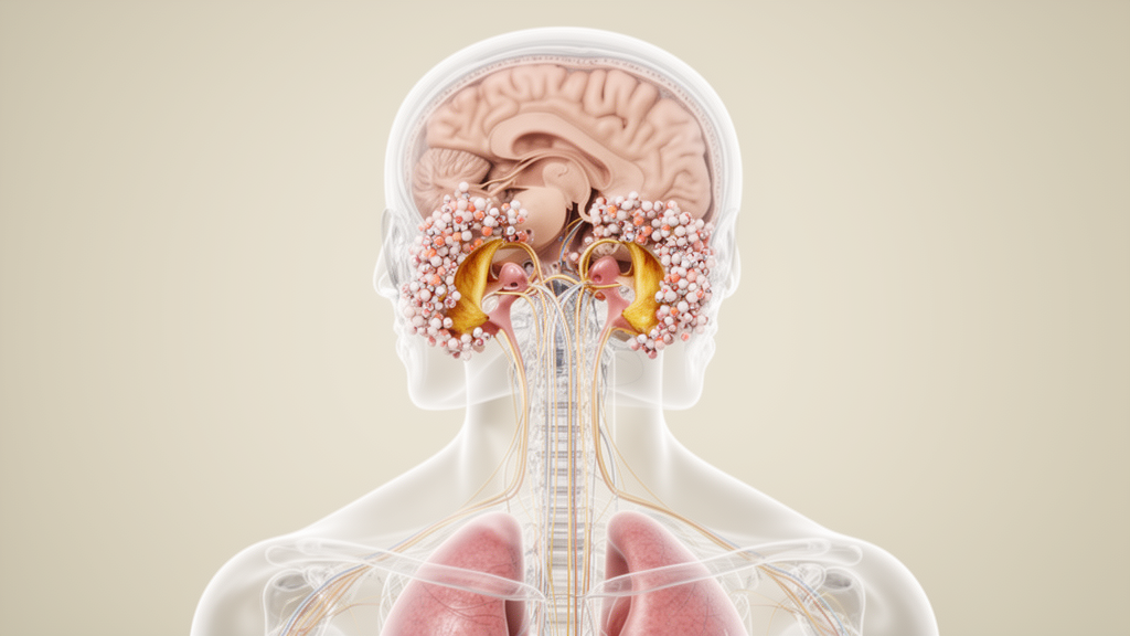
The case report found that cancer cells were present in the cerebrospinal fluid and that magnetic resonance imaging showed enhancement along the leptomeninges and around both vestibulocochlear nerves. Quantitative video head impulse testing documented impaired vestibular function in all six semicircular canals. Treatment with intrathecal pemetrexed and dexamethasone was followed by improvement in dizziness and no discernible tumor cells in subsequent cerebrospinal fluid samples, although the patient died about one year after discharge as his malignancy progressed.
The patient’s symptoms had begun six months before hospital admission. He described dizziness, recurrent tinnitus, oscillopsia, imbalance and bilateral hearing loss, with his balance becoming worse when standing, moving in darkness and walking at night. He had lived with lung adenocarcinoma for more than a decade and was receiving osimertinib at 160 mg once daily.
He reported no fever, ear pain or discharge. There was also no history of vestibulotoxic drug administration, and the investigators did not consider the targeted cancer therapy to be the cause of his vestibular symptoms because they began four years after treatment started and clinical trials had reported no ototoxic effects. Examination revealed a suspicious positive clinical horizontal head impulse test on the left, bilateral reduction of hearing to finger rub, and positive Romberg and tandem-Romberg tests.
There was no spontaneous nystagmus, and the remaining neurological examination did not reveal facial palsy, limb weakness, sensory loss, ataxia, pyramidal signs or meningeal irritation. Otoscopy found patent external auditory canals and intact tympanic membranes without discharge or signs of active infection. An ear, nose and throat consultation found no evidence of Ménière’s disease, autoimmune inner-ear disease or labyrinthitis.
Quantitative testing supplied more specific evidence of the extent of the vestibular injury. A high-frequency video head impulse test, or vHIT, showed reduced vestibulo-ocular reflex gain in all six semicircular canals, covering the horizontal and vertical canals of both inner ears. Audiometry demonstrated bilateral sensorineural hearing loss that was more severe on the right, while brainstem-evoked auditory potentials were normal.
Plain CT and MRI scans of the cochleae and cochlear nerves were unremarkable, providing no evidence of a destructive inner-ear lesion on those images. The combination of bilateral sensorineural hearing loss and reduced vestibular responses established that the patient had dysfunction of both the auditory and vestibular sides of the peripheral nervous system. Contrast-enhanced brain MRI revealed enhancement of both vestibulocochlear nerves and along the cerebral leptomeninges.
Spinal MRI identified metastases at T10, L1 and L4. Chest CT showed a 22 × 13 mm mass-like high-density lesion in the anterior segment of the left upper lobe, which was considered suggestive of a tumor. The broader imaging pattern showed that the vestibular and auditory deficits were not confined to the inner ears but formed part of a metastatic process involving the meninges, cranial nerves and spine.
Bilateral vestibulopathy can arise from several different causes, including ototoxic drugs, bilateral Ménière’s disease, meningitis and genetic disorders. The report noted that definite causes were identified in approximately 30% of one 255-patient cohort and 47% of a separate 154-patient cohort, leaving a substantial proportion of cases without an established etiology. Diagnostic lumbar puncture added further evidence.
The cerebrospinal fluid had normal pressure, glucose and chloride concentrations, but protein was elevated to 110.2 mg/dL and IgG to 144 mg/L. It also contained 44 nucleated cells per microliter, with 84% lymphocytes, and cytology revealed individually dispersed cells with nuclear atypia and degeneration. Serum testing was positive for anti-AQP4 IgG at a titer of 1:320, introducing the possibility of a paraneoplastic or autoimmune process.
However, none of the other 30 paraneoplastic antibodies tested was positive, including KLHL11-IgG, which is associated with bilateral vestibular dysfunction, and ANNA-1, or anti-Hu. The patient also lacked the core clinical syndromes associated with AQP4-IgG-positive neuromyelitis optica spectrum disorder, including optic neuritis, acute myelitis, area postrema syndrome and characteristic brainstem, diencephalic or cerebral syndromes. Using the PNS-Care scoring system, the researchers classified the presentation as a possible, rather than confirmed, paraneoplastic neurologic syndrome.
The four points came from the presence of cancer; anti-AQP4-IgG did not add points because it was classified as a lower-risk antibody. The authors therefore judged direct metastatic infiltration to be the best-supported explanation. They based that conclusion on malignant cells in the cerebrospinal fluid, leptomeningeal enhancement, bilateral vestibulocochlear nerve enhancement and the absence of alternative inflammatory or autoimmune manifestations.
Nerve enhancement alone would not have distinguished leptomeningeal metastasis from paraneoplastic cochleovestibulopathy: internal auditory canal enhancement was reported in 4 of 26 patients, or 15%, with paraneoplastic cochleovestibulopathy, while leptomeningeal disease can initially appear as isolated bilateral vestibulocochlear nerve involvement. In the present case, the additional leptomeningeal abnormalities and malignant cerebrospinal fluid findings favored metastatic disease. There was also an anatomical explanation consistent with the observed pattern.
Metastatic cells can spread through cerebrospinal fluid and seed sites where flow is slow or where gravity and fluid absorption influence deposition. The vestibular nerve exits the brainstem at the cerebellopontine angle and passes through an area favorable to meningeal seeding. Because the vestibular and auditory nerves are wrapped in pia mater and traverse regions containing cerebrospinal fluid, metastatic cells can encounter and damage these nerves directly.
In this patient, the same cerebrospinal fluid that carried malignant cells also bathed both affected nerves. The pattern of imaging, cytology and objective vestibular testing therefore supported direct metastatic seeding rather than a remote immune-mediated paraneoplastic effect, although the report described this as the pathophysiologic mechanism most consistent with the available evidence. After discharge, the treatment strategy was changed to intrathecal pemetrexed at 15 mg plus dexamethasone at 5 mg when the initial furmonertinib treatment proved insufficient.
The patient underwent several treatment cycles. His dizziness subsequently subsided, and repeat cerebrospinal fluid analysis found no discernible tumor cells, changes that occurred alongside his symptomatic improvement. The investigators could not repeat vHIT because treatment and follow-up occurred across specialized departments at other hospitals.
They identified six earlier cases with objectively documented bilateral vestibular dysfunction, all individual case reports: three had been assessed with caloric testing, one with rotary harmonic stimulation combined with electronystagmography, one with electronystagmography and dynamic posturography, and one with caloric testing followed by a bedside head impulse test. An additional 23 publications described auditory or vestibular symptoms associated with leptomeningeal metastasis but did not provide objective evidence of bilateral vestibular hypofunction. The present vHIT assessment was therefore distinct from the methods used in those earlier reports.
Several limitations constrain what the case can establish. Symptom information during follow-up was obtained by telephone and from family rather than direct clinical examination. vHIT was performed only once, no follow-up brain or spinal MRI was obtained, and no specific treatment was documented for the vertebral metastases. The patient later declined further antitumor treatment and died approximately one year after discharge following clinical deterioration attributed to progressive malignancy.
Consequently, the report cannot determine whether the neurological improvement was durable, whether vestibular-test gains recovered, or whether imaging changes accompanied the clinical improvement. As a single case with a limited treatment literature, it cannot establish comparative effectiveness or prognosis for a broader population. Its central finding is that bilateral vestibulopathy, accompanied by hearing loss and confirmed impairment of all six semicircular canals, was documented as an uncommon manifestation of leptomeningeal metastasis from lung adenocarcinoma.
The authors emphasized that the combination of contrast-enhanced MRI and cerebrospinal fluid cytology identified the malignant cause in this patient, while the positive anti-AQP4-IgG result was treated as an accompanying serological finding rather than evidence of neuromyelitis optica spectrum disorder or a paraneoplastic mechanism.
Subject of Research: Lung adenocarcinoma-associated leptomeningeal metastasis causing bilateral vestibulopathy
Article Title: Case Report: Bilateral vestibulopathy associated with leptomeningeal metastasis
Article References: Feng, D., Wei, W., Hu, S., & Hong, X. (2026). Case Report: Bilateral vestibulopathy associated with leptomeningeal metastasis. Frontiers in Oncology, 16, Article 1981680. https://doi.org/10.3389/fonc.2026.1981680
Image Credits: AI Generated
DOI: 10.3389/fonc.2026.1981680
Keywords: leptomeningeal metastasis, bilateral vestibulopathy, lung adenocarcinoma, vestibulocochlear nerves, video head impulse test, cerebrospinal fluid cytology, sensorineural hearing loss, oscillopsia, intrathecal chemotherapy, case report, Case, Report
Cite Scienmag News
Nathaniel Bowman. (October 20, 2026). Case report links lung cancer leptomeningeal metastasis to bilateral vestibulopathy. Scienmag. https://scienmag.com/case-report-links-lung-cancer-leptomeningeal-metastasis-to-bilateral-vestibulopathy/
Nathaniel Bowman. "Case report links lung cancer leptomeningeal metastasis to bilateral vestibulopathy." Scienmag, 20 October 2026, https://scienmag.com/case-report-links-lung-cancer-leptomeningeal-metastasis-to-bilateral-vestibulopathy/. Accessed 20 October 2026.
Nathaniel Bowman. "Case report links lung cancer leptomeningeal metastasis to bilateral vestibulopathy." Scienmag. October 20, 2026. https://scienmag.com/case-report-links-lung-cancer-leptomeningeal-metastasis-to-bilateral-vestibulopathy/


