


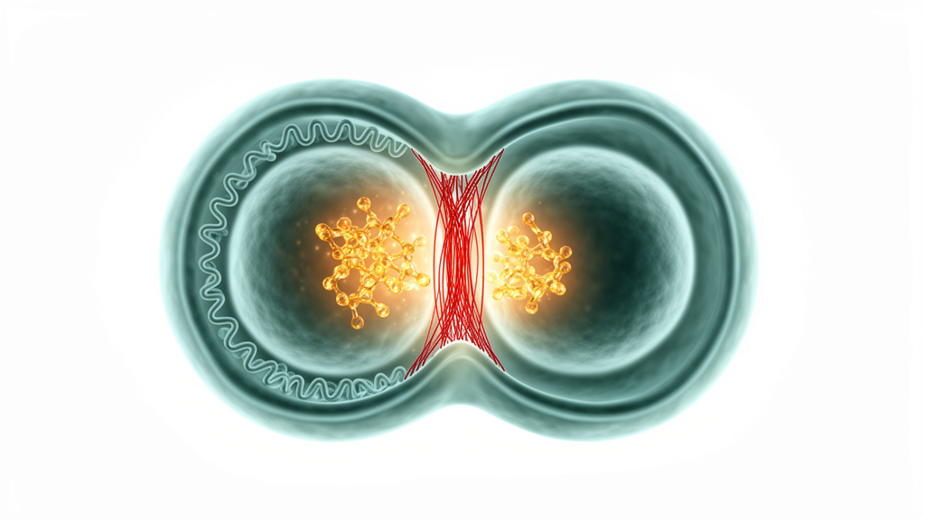
For decades, AXIN1 has been a familiar name in cell biology laboratories, celebrated as the scaffold protein that holds together the β-catenin destruction complex alongside APC and GSK3β, thereby governing the canonical Wnt signaling pathway. Its ubiquitously expressed cousin AXIN2, by contrast, behaves as a Wnt target gene whose levels rise in step with β-catenin accumulation. AXIN1 has also been implicated in TGF-β signaling as an adaptor for SMAD3, in the SAPK/JNK pathway where its overexpression triggers apoptosis through MEKK activation, and in p53 signaling following DNA damage. Cancer cells frequently carry AXIN1 mutations that confer resistance to apoptosis, and restoring AXIN1 expression in hepatocellular and colon cancer cells can induce cell death in a JNK-dependent manner. Yet a new study published in iScience by Na Gyeong Lee, Na Yeong Lee, Mi-Lang Kyun, Ji Eun Yu, Sun-Ok Kim, Yoon-Hee Na, Taeho Kwon, Hee Jun Cho, Key-Hwan Lim, Kyung Ho Lee and colleagues reveals that this versatile scaffold protein has been quietly moonlighting in one of the most dramatic moments of the cell cycle: the physical split of one cell into two.
The research team, working with HeLa CCL2 and MCF-7 cells, discovered that AXIN1 accumulates in the midzone during anaphase and localizes specifically to the midbody in telophase cells, a localization that had never been reported before. Using gentle detergent extraction with 0.2% Triton X-100 before fixation, followed by immunostaining with two distinct anti-AXIN1 antibodies, the investigators confirmed this unexpected midbody enrichment. Co-staining with membrane markers such as Na+/K+-ATPase and E-CADHERIN further validated the finding. The midbody, sometimes called the Flemming body, telophase disk, or stem body, is the phase-dense structure that forms at the center of the narrow intercellular bridge connecting two sister cells during the final stages of division. Its completion ultimately results in two independent daughter cells, and the process of cytokinesis unfolds through four sequential stages: positioning of the cleavage plane, cleavage furrow ingression, midbody formation, and abscission.
What makes this discovery particularly striking is how AXIN1’s midbody localization dovetails with the team’s previous work on β-catenin. In an earlier study, the researchers had shown that phosphorylation of β-catenin at Ser60 by polo-like kinase 1 (PLK1) in early telophase cells recruits ECT2 to the midbody, which in turn activates RHOA to drive cleavage furrow ingression and complete cytokinesis. Now, the team found that AXIN1 colocalizes with β-catenin p-S60 at the midbody during early telophase, and immunoprecipitation analysis revealed a significant increase in the interaction of AXIN1 with β-catenin, β-catenin p-S60, PLK1, ANILLIN, and ECT2 during mitosis. These results suggest that AXIN1 shares several functions with β-catenin p-S60 during mitosis and that the β-catenin p-S60-dependent cytokinetic components likely involve AXIN1 during early telophase.
To test whether AXIN1 is genuinely required for this machinery, the researchers knocked down endogenous AXIN1 using two different siRNAs, si778 and si873. The consequences were dramatic. Depletion of AXIN1 resulted in a marked absence of ECT2 at the midbody in early telophase cells, and RHOA activation assays showed that the level of active RHOA in lysates from AXIN1-depleted cells was substantially decreased compared with control knockdown cells. Immunostaining with an anti-α-tubulin antibody confirmed that the frequency of multinucleated cells was significantly increased by AXIN1 depletion, with an approximately 8-fold increase using si778 and a 4-fold increase using si873 compared with the control. The intercellular bridge phenotype also increased roughly 2-fold. Fluorescence-activated cell sorting analysis revealed an increase in >4N cells and a slight increase in sub-G1 cells in AXIN1 knockdown populations, and KI67 staining showed an overall reduction in proliferative activity, indicating that AXIN1 loss impairs proper cell-cycle progression and cell division.
Live-cell imaging provided perhaps the most vivid evidence of AXIN1’s importance. Time-lapse video analysis revealed that 92% of cells transfected with control siRNA underwent normal division and successful abscission, with only 8% showing a delayed process. In stark contrast, knocking down AXIN1 significantly amplified the rate of division errors, with 16% of cells displaying delayed cytokinesis and 61% experiencing regression after cleavage furrow ingression. Numerous cells lacking AXIN1 could not finalize division after furrowing, ultimately leading to regression, sometimes accompanied by severe chromosome segregation defects. These time-lapse results establish that the absence of AXIN1 increases the frequency of cytokinetic delays and regression following furrow formation, painting a picture of a scaffold protein whose loss sends the delicate choreography of cell division spinning into chaos.
But AXIN1’s story at the midbody does not end with furrow ingression. Super-resolution microscopy using a Zeiss LSM-880 equipped with Airyscan confirmed that AXIN1 forms a ring-like structure in the Flemming body that completely co-localizes with the ring-like structures of CEP55 and MKLP1, both known midbody ring components. Co-immunostaining with a panel of midbody markers, including ANILLIN, ECT2, INCENP, Aurora B, KIF4A, PRC1, CHMP4B, ALIX, ACTIN, and α-tubulin, further highlighted AXIN1’s specific localization to the Flemming body and dark zone within the intercellular bridge. Immunoprecipitation analysis in cells released at 2 hours after nocodazole trapping revealed that endogenous AXIN1 was complexed with CEP55 or MKLP1 in anaphase and telophase cell populations, suggesting that AXIN1 functions as a midbody ring component during late telophase.
This ring-like localization led the team to investigate whether AXIN1 participates in the abscission machinery, the final stage of cytokinesis during which the cytoplasmic bridge contracts to a diameter of approximately 0.2 micrometers and the microtubule bundles disintegrate. The endosomal sorting complex required for transport, or ESCRT, plays a crucial role at this stage, working sequentially through four groups of machinery: ESCRT 0, ESCRT I, ESCRT II, and ESCRT III. The researchers found that AXIN1 is involved in the midbody localization of several abscission factors, including CEP55, ALIX, TSG101, and CHMP4B, but not MKLP1. Following AXIN1 knockdown, the CEP55 signal intensity in the midbody was reduced by approximately 75% compared with control cells, ALIX signal intensity dropped by roughly 75%, and the midbody signals for TSG101 and CHMP4B were reduced by approximately 50%.
Intriguingly, the midbody localization of MKLP1, a central spindle-associated protein, was not reduced by AXIN1 knockdown but was instead enhanced. This discrepancy may result from the disruption of the sequential recruitment of the centralspindlin and abscission components to the midbody, excluding MKLP1. To determine where AXIN1 sits in this hierarchy, the team depleted MKLP1 and examined AXIN1’s midbody localization. The depletion of MKLP1 reduced the midbody localization of AXIN1 by approximately 50% compared with control knockdown cells. These findings suggest that AXIN1 is localized between MKLP1 and CEP55, thereby promoting the recruitment of ESCRT components such as ALIX, TSG101, and CHMP4B to the midbody, and establishing an MKLP1-AXIN1-CEP55-ALIX-TSG101-CHMP4B abscission axis.
The study also uncovered a curious biochemical detail about how AXIN1 engages with PLK1, the kinase that orchestrates so many mitotic events. AXIN1 binds to the PLK1 polo-box domain (PBD) in a phosphorylation-independent manner, as demonstrated by GST-PBD binding assays using three different cell states: asynchronously growing cells, thymidine-treated S-phase cells, and nocodazole-treated G2/M-phase cells. Similar amounts of AXIN1 were bound between PLK1 PBD wild-type and the H538A/K540M mutant at all cell stages examined, unlike other PLK1 substrates that require priming phosphorylation. This mirrors the previously reported pattern of β-catenin binding to the PLK1 PBD during cytokinesis, also in a phosphorylation-independent manner, and suggests that AXIN1 and PLK1 may function together during both interphase and mitosis without the conventional phosphodependent substrate targeting that characterizes most PLK1 interactions.
The implications of this dual-function discovery extend well beyond basic cell biology. AXIN1 deficiency and mutation are well established contributors to carcinogenesis through abnormalities in Wnt signaling, and defects in cytokinesis that lead to genomic instability and tumorigenesis have also been reported. The researchers propose that AXIN1 acts as a cytokinetic factor participating from early to late telophase, first as a component of the β-catenin p-S60-dependent cytokinesis machinery during early telophase, and second as a component of the MKLP1-AXIN1-CEP55-ALIX-TSG101-CHMP4B abscission axis in late telophase. Whether the oncogenic potential of AXIN1 dysregulation leads to abnormal cytokinesis, and whether the oncogenic mechanism of AXIN1 dysfunction in canonical Wnt signaling is interrelated or independent of its scaffolding role in cytokinesis, remains an open question. The authors acknowledge that the exact mechanisms distinguishing AXIN1’s stage-specific functions remain unsolved, and future studies must investigate the temporal changes in the AXIN1 protein, the upstream factors driving these modifications, and the dynamic network of co-regulatory interacting factors during each cytokinetic stage. What is clear, however, is that this unassuming scaffold protein, long studied for its role in deciding the fate of β-catenin, has now been caught red-handed at the very center of the cell’s final act of division, holding together the molecular machinery that allows one cell to become two.
Subject of Research: Dual roles of AXIN1 as a β-catenin p-S60-dependent cytokinesis machinery and an ESCRT component
Article Title: Dual roles of AXIN1 as a β-catenin p-S60-dependent cytokinesis machinery and an ESCRT component
Article References: Lee, N. G., Lee, N. Y., Kyun, M.-L., Yu, J. E., Kim, S.-O., Na, Y.-H., Kwon, T., Cho, H. J., Lim, K.-H., & Lee, K. H. (2026). Dual roles of AXIN1 as a β-catenin p-S60-dependent cytokinesis machinery and an ESCRT component. iScience, 29(11), Article 117923. https://doi.org/10.1016/j.isci.2026.117923
Image Credits: AI Generated
DOI: 10.1016/j.isci.2026.117923
Keywords: Dual, roles, AXIN1, catenin, p-S60-dependent, cytokinesis, machinery, ESCRT, component, scientific research
Cite Scienmag News
Denise Maddox. (October 18, 2026). AXIN1 Found Pulling Double Duty in Wnt Signaling and Cytokinesis. Scienmag. https://scienmag.com/axin1-found-pulling-double-duty-in-wnt-signaling-and-cytokinesis/
Denise Maddox. "AXIN1 Found Pulling Double Duty in Wnt Signaling and Cytokinesis." Scienmag, 18 October 2026, https://scienmag.com/axin1-found-pulling-double-duty-in-wnt-signaling-and-cytokinesis/. Accessed 18 October 2026.
Denise Maddox. "AXIN1 Found Pulling Double Duty in Wnt Signaling and Cytokinesis." Scienmag. October 18, 2026. https://scienmag.com/axin1-found-pulling-double-duty-in-wnt-signaling-and-cytokinesis/


