


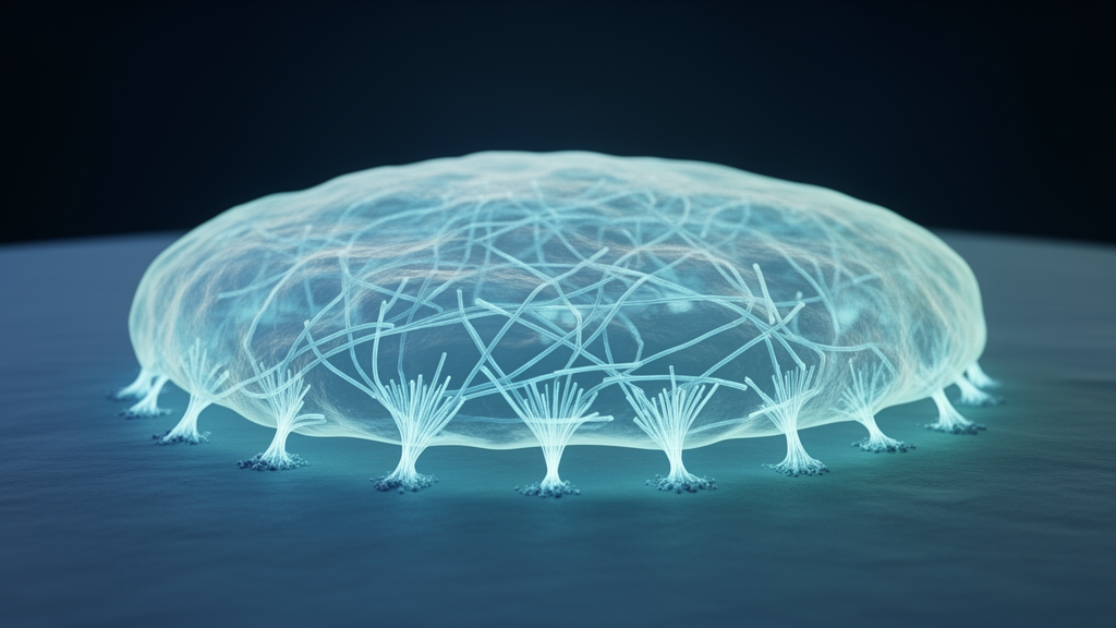
Every cell in the body lives surrounded by mechanical information. The stiffness of the tissue it occupies, the pull of neighboring cells, the stretch of a beating heart or the flow of fluid along a blood vessel: all of these forces are translated into biochemical signals that ultimately reshape the cell from within. A new review published in Molecular Biology Reports by Gangotry Mukhopadhyay, Karthick Velu, Inbakandan Dhinakarasamy, Prathap Somu, J. Iyyappan, Ravi Mani and colleagues brings together the rapidly expanding field of adhesion mechanobiology into a single framework, arguing that cellular adhesion is the central gateway through which mechanical cues trigger cytoskeletal reconfiguration. The work arrives at a moment when tissue engineering, cultivated meat production, cancer research and regenerative medicine all depend on the same fundamental question: how can dissociated cells be coaxed into forming stable, functional three-dimensional structures without losing their integrity?
The review’s central premise is that adhesion is not a passive glue but an active signaling hub. When a cell contacts the extracellular matrix, transmembrane receptors called integrins bind to motifs such as the arginine-glycine-aspartate, or RGD, sequence found in fibronectin and other matrix proteins. This binding event nucleates a focal adhesion, a nanoscale assembly of more than a hundred proteins including talin, vinculin, paxillin, kindlin and focal adhesion kinase. Talin acts as the critical mechanical link, stretching under force to expose binding sites for actin filaments of the cytoskeleton. Vinculin reinforces this linkage, and the review highlights recent work measuring the mechanical response of vinculin directly, showing how the protein unfolds and refolds under tension to tune adhesion strength. The result is a molecular clutch that connects the outside world to the cell’s internal scaffold.
What makes focal adhesions remarkable is their layered architecture. Cited work in the review describes a stratified organization in which integrins at the membrane face, adapter proteins in the intermediate zone and actin regulators at the deepest layer transmit force across tens of nanometers with remarkable fidelity. Single-molecule tension sensors, including DNA-based probes and plasmonic nanosensors, have now revealed that individual adhesion bonds experience piconewton-scale forces that fluctuate over milliseconds. A 2026 study cited by the authors linked molecular tension to cellular traction forces in a multiscale analysis of focal adhesion mechanics, confirming that the forces cells exert on their surroundings are built up from the sum of these molecular-scale events. Computational models of force-dependent adhesion assembly and disassembly further show how adhesions can self-stabilize under load, a generic mechanism that helps cells maintain grip in a mechanically noisy environment.
Downstream of the adhesion complex, mechanical signals cascade into the cytoskeleton and ultimately into the nucleus. The Rho family of small GTPases, acting through effectors such as Rho-associated protein kinase, or ROCK, and the beta-PIX/GIT complex, remodels actin filaments in response to adhesion cues. Focal adhesion kinase activates signaling pathways including JNK, which the review notes is activated by mechanical stress in an integrin-dependent manner. One of the most consequential endpoints is the transcriptional co-activator YAP, the nuclear messenger of the Hippo pathway. Recent work cited in the review demonstrates that FAK, vinculin and talin jointly control mechanosensitive YAP nuclear localization, meaning that the physical state of adhesions directly determines which genes the cell expresses. In endothelial cells, substrate stiffness modulates function through a YAP-Dll4-Notch1 pathway, illustrating how mechanical information flows all the way from the matrix to developmental signaling.
The review devotes substantial attention to substrate stiffness, one of the most influential discoveries in modern cell biology. The landmark finding that matrix elasticity directs stem cell lineage specification, published nearly two decades ago, showed that mesenchymal stem cells become bone-like on stiff substrates and brain-like on soft ones, purely as a function of the elasticity beneath them. The new review extends this picture with recent evidence that substrate stiffness alters the layer architecture and biophysics of human induced pluripotent stem cells, modulating their differentiation potential. It also complicates the story: polyacrylamide hydrogels with rigidity-independent surface chemistry show limited long-term maintenance of pluripotency on soft substrates, suggesting that stiffness sensing and surface chemistry interact in ways that are still being untangled. Cardiomyocytes, fibroblasts and cancer cell lines each respond to stiffness gradients in distinct ways, and the review emphasizes that some stiffness sensing by fibroblasts is myosin-independent, regulated instead by the viscoelasticity of flowing actin networks.
Beyond stiffness, the authors catalog the experimental toolbox that mechanobiologists now use to interrogate adhesion. Micropatterning techniques constrain cell shape and ligand spacing on engineered surfaces, and studies cited in the review show that cell shape and tension alone can alter focal adhesion structure, while biomimetic micropatterned surfaces change the adhesion and morphology of cervical cancer cells. Titanium dioxide nano-biopatterning has revealed optimal ligand presentation for cell-matrix adhesion formation. Stretch devices apply cyclic strain to cells grown on flexible membranes, and dimensionality matters: mechanical stretch regulates cell behavior differently in two-dimensional culture than in three-dimensional hydrogels. Recent work on cell-matrix feedback shows that stretch-induced cellular memory and fibroblast activation depend on the mechanical properties of the matrix itself, hinting that cells remember the forces they have experienced through persistent changes in their adhesion machinery.
Chemical strategies complete the toolkit. Thermoresponsive polymers allow reversible cell adhesion that can be switched on and off by temperature, a property exploited in cell sheet engineering. Reversible host-guest interactions in three-dimensional microscaffolds permit mechanical stimulation of single cells with exquisite spatial control, and photo-switchable surfaces enable spatiotemporal modulation of cytoskeletal forces that visibly deform the nucleus. Amphiphilic cationic cyclodextrin nanovesicles serve as versatile cues for guiding cell adhesion, and strain-dependent glutathionylation of fibronectin fibers has been shown to prime an integrin switch, a striking example of chemistry and mechanics coupling within the matrix itself. Even the extracellular domains of E-cadherin, the prototypical cell-to-cell adhesion molecule, determine mechanical phenotypes of epithelia through outside-in signaling, underscoring that the review’s framework spans both cell-matrix and cell-cell junctions, where cadherins and F-actin engage in a positive feedback loop initiated by cortical tension.
Measurement technology occupies a special place in the review, particularly bio-atomic force microscopy. AFM can map the local stiffness of living cells, probe adhesion forces with piconewton sensitivity and, in newer modes such as nanoendoscopy, visualize intracellular nanostructures in living cells. Fluidic force microscopy allows simultaneous measurement of single-cell mechanics and adhesion to materials, while atomic force acoustic microscopy extends mechanobiological analysis to manifold live cell types in vitro. The integration of AFM with fluorescence microscopy, including FRET-based tension probes and super-resolution imaging of DNA-based tension sensors, is transforming the field’s ability to watch mechanotransduction happen in real time. Deep learning-assisted AFM image recognition now enables efficient single-cell mechanics measurements even in co-culture environments, and roughness-driven compressive sensing improves nanoscale surface characterization. The review positions these quantitative methodologies as the connective tissue of adhesion mechanobiology, the tools that turn qualitative observations into testable, predictive models.
The applications flowing from this unified framework are broad. In tissue engineering, scaffold-free three-dimensional-printed heart tissue grafts develop better under mechanical stimulation, and micropatterned nanofiber scaffolds guide muscle tissue formation. Biomechanical stimulation of mesenchymal stem cells in three-dimensional peptide nanofibers promotes bone differentiation. In cancer biology, adhesion mechanics govern how tumor cells migrate through constrained microenvironments, and engineered T cells are being designed to navigate the structurally complex terrain of tumors. Adhesion signaling is also entwined with cellular senescence, with integrin-mediated adhesions regulating the entry of cells into aged, dysfunctional states, a connection with implications for age-related diseases such as emphysema. By integrating mechanical stimulation strategies, chemotransduction, adhesion-associated molecular networks and AFM-based quantification into one coherent picture, the review makes a compelling case that the adhesion gateway is where biology meets physics, and that learning to speak its language will define the next generation of therapies and engineered tissues.
Subject of Research: Mechanobiological signaling at cell adhesion sites and its role in cytoskeletal reconfiguration
Article Title: Adhesion gateway: decoding mechanobiological cues for cytoskeletal reconfiguration
Article References: Mukhopadhyay, G., Velu, K., Dhinakarasamy, I., Somu, P., Iyyappan, J., & Mani, R. (2026). Adhesion gateway: decoding mechanobiological cues for cytoskeletal reconfiguration. Molecular Biology Reports, 53(1), Article 1756. https://doi.org/10.1007/s11033-026-12912-6
Image Credits: AI Generated
DOI: 10.1007/s11033-026-12912-6
Keywords: mechanobiology, focal adhesion, integrins, cytoskeleton, mechanotransduction, talin, vinculin, YAP, substrate stiffness, atomic force microscopy, tissue engineering, cell adhesion
Cite Scienmag News
Drew Townsend. (October 17, 2026). Cells Take the Hint: How Adhesion Gateways Reshape the Cytoskeleton. Scienmag. https://scienmag.com/cells-take-the-hint-how-adhesion-gateways-reshape-the-cytoskeleton/
Drew Townsend. "Cells Take the Hint: How Adhesion Gateways Reshape the Cytoskeleton." Scienmag, 17 October 2026, https://scienmag.com/cells-take-the-hint-how-adhesion-gateways-reshape-the-cytoskeleton/. Accessed 17 October 2026.
Drew Townsend. "Cells Take the Hint: How Adhesion Gateways Reshape the Cytoskeleton." Scienmag. October 17, 2026. https://scienmag.com/cells-take-the-hint-how-adhesion-gateways-reshape-the-cytoskeleton/


