


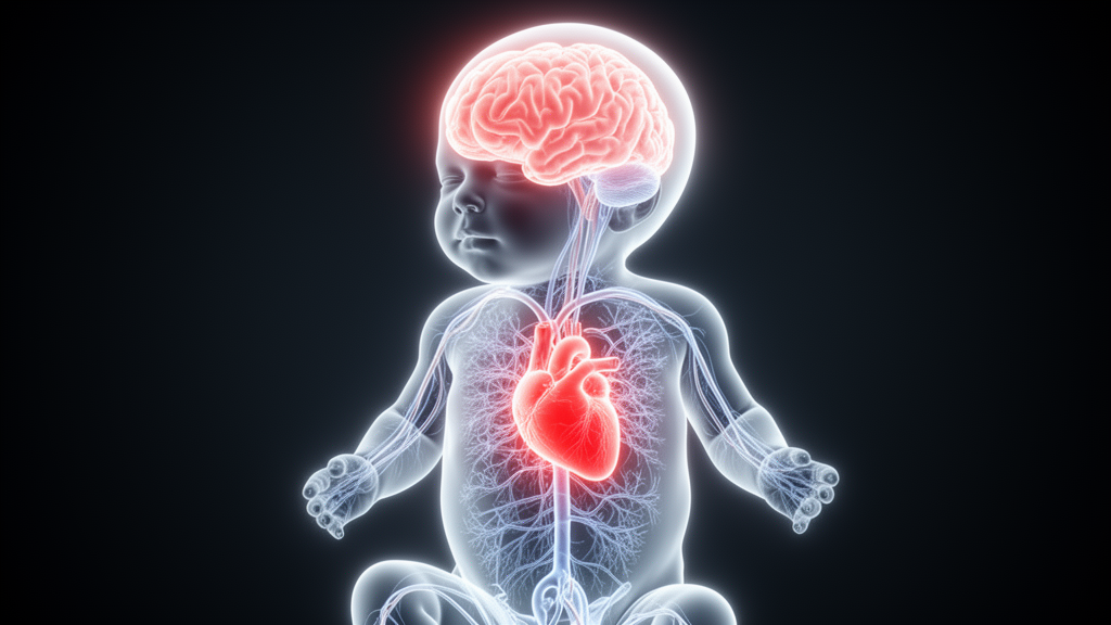
Every minute of a newborn’s life, two of the body’s most vital systems—the brain and the heart—are engaged in a silent, continuous dialogue. Electrical storms of cortical activity ripple through the infant’s developing brain, while the heart responds with subtle shifts in its rhythm. Now, a team of Ukrainian researchers has found a way to eavesdrop on that conversation, and what they heard may change how doctors detect one of the most devastating conditions in neonatal medicine: hypoxic-ischemic encephalopathy, or HIE, a form of brain injury caused by oxygen deprivation around the time of birth.
HIE is a leading cause of neonatal death and lifelong neurological disability worldwide. When a baby’s brain is starved of oxygen and blood flow during the perinatal period, the consequences can be catastrophic—but the window for intervention is heartbreakingly narrow. The gold standard for assessing severity, the Sarnat staging system introduced in 1976, classifies infants into three clinical stages based on observable signs: Stage 1 (mild) presents with hyper-alertness and irritability, Stage 2 (moderate) involves lethargy and suppressed reflexes, and Stage 3 (severe) manifests as profound coma. The problem is that the transition from mild to moderate injury—the exact moment when therapeutic hypothermia becomes critical—is often clinically ambiguous, with symptoms that overlap and subjective assessments that vary between observers.
Now, a pilot study published in Frontiers in Network Physiology has taken a fundamentally different approach. Rather than looking at the brain and heart in isolation, researchers led by Danylo Reznichenko of Igor Sikorsky Kyiv Polytechnic Institute and colleagues at Bukovinian State Medical University asked a deceptively simple question: what happens to the conversation between these two organs when the brain is injured? Their answer, drawn from synchronized long-term EEG and ECG recordings of 61 neonates, is that the dialogue doesn’t just fade—it reorganizes in ways that are quantifiable, measurable, and potentially diagnostic.
The team collected data from the Neonatal Intensive Care Unit of the Central Municipal Clinical Hospital in Chernivtsi, Ukraine, recording continuous EEG from two channels (F3-P3 and F4-P4) alongside ECG from the C3-C4 position. The recordings, captured in standard BioSemi format using a clinical-grade bedside monitor, ranged from 19 minutes to over 13 hours, preserving the full macro-scale dynamics of sleep-wake cycling and metabolic drift. The core analytical cohort focused on term and near-term neonates with gestational ages between 34 and 41 weeks—a deliberate constraint that isolates the analysis from the confounding effects of gestational immaturity, since infants in this window exhibit fully established baseline electrocortical patterns.
The technical pipeline was formidable. The researchers extracted a staggering 774 distinct features across five physiological domains: 200 from amplitude-integrated EEG statistics, 40 from quantitative EEG spectral analysis, 132 from heart rate variability metrics, 130 from ECG statistical and spectral properties, and 272 from fine-grained cardiocycle morphological descriptors tracking the P, Q, R, S, and T waveforms of individual heartbeats. The amplitude-integrated EEG was generated using the established algorithm of Zhang and Ding, with asymmetric bandpass filtering between 0.5 and 15 Hz, amplitude envelope extraction via the Hilbert transform, and semi-logarithmic amplitude compression. Heart rate variability features were derived using the pyHRV library, with ectopic beat detection and outlier correction protocols built in, while ECG signals were cleaned using NeuroKit2’s two-stage pipeline—a fourth-order Butterworth high-pass filter at 0.5 Hz to eliminate baseline wander, followed by a 50 Hz notch filter to suppress powerline interference.
The central finding is stark. Neonates with healthy continuous normal voltage (CNV) EEG patterns exhibited robust neuro-cardiac synchronization, with a median Spearman correlation coefficient of 0.22 between the aEEG envelope and heart rate dynamics. But in infants with abnormal EEG backgrounds—burst suppression, continuous low voltage, or discontinuous normal voltage—that synchronization collapsed by approximately 60 percent, dropping to a median of 0.09 (p=0.015). This isn’t merely a statistical curiosity; it suggests that electrocortical integrity is a fundamental prerequisite for the flexible, responsive dialogue between brain and heart, and that its breakdown serves as a quantifiable marker of neurological compromise.
Even more intriguing is what the temporal lag analysis revealed. Using constrained cross-correlation between the smoothed aEEG upper envelope and the continuous heart rate trace within 300-second sliding windows, the researchers found that cardiovascular fluctuations typically preceded cortical envelope changes in Stage 1 neonates, with a median negative lag of −2.08 seconds. This
The physiological logic behind this brain-heart dialogue rests on the autonomic nervous system, the bidirectional communication network that links the central nervous system to the heart through sympathetic and parasympathetic pathways. In healthy newborns, heart rate variability—the beat-to-beat fluctuation in the intervals between successive heartbeats—reflects a mature balance between these two branches. The sympathetic arm prepares the body for stress and accelerates the heart, while the parasympathetic arm, mediated largely by the vagus nerve, slows it down. When the brain sustains hypoxic-ischemic injury, this autonomic regulation is often among the earliest casualties, which is precisely why heart rate variability has long been investigated as a potential window onto neurological status in the neonatal intensive care unit.
Earlier work has consistently pointed in this direction. Studies of preterm infants have shown that bursts of slow-wave activity on the EEG are synchronized with transient accelerations of heart rate, suggesting that even in the immature brain, cortical events and cardiac responses are temporally locked. In neonates experiencing seizures—a frequent complication of HIE—researchers have documented significant correlations between instantaneous heart rate changes and EEG amplitude, with some analyses finding that the coupling appeared in every patient studied and in the majority of individual seizure events. Adding ECG channels to conventional EEG monitoring has even been shown to improve seizure detection rates substantially, from roughly 81 percent with EEG alone to 98 percent when both signals were combined in one cohort of term infants.
Heart rate variability metrics themselves have proven informative in HIE. Time-domain measures such as the standard deviation of normal-to-normal intervals and frequency-domain components in the low-frequency and high-frequency bands all tend to decrease as EEG background abnormalities worsen, with reported correlations reaching moderately strong values. Other investigators have combined HRV features spanning time-domain, frequency-domain, and entropy measures with clinical data to predict EEG severity grade, achieving respectable discrimination in classification tasks. These converging lines of evidence established the biological plausibility of the coupling approach before the current study quantified it directly.
The conceptual framing of the research draws on Network Physiology, an emerging discipline that treats the human organism as an integrated network of interacting organ systems rather than a collection of independently functioning parts. Within this paradigm, health is characterized by flexible, multi-scale coordination among subsystems, while disease manifests as a disruption of that coordination—either a rigidification of interactions or a fragmentation of the network’s topology. The brain-heart axis is one of the most intensively studied edges in this network, and neonatal HIE offers a natural experiment in how a systemic insult can reorganize cross-organ dynamics in a measurable way.
The machine learning component of the study deserves particular attention for its methodological caution. In applied machine learning, it is easy to report a single best-performing configuration and present it as the capability of a system, but such peak values often reflect overfitting to particular data partitions or lucky hyperparameter draws. By evaluating models across diverse parameter grids and reporting average performance across window lengths alongside maximum values, the researchers provided a more honest picture of what automated HIE grading can currently achieve. The gap between the peak accuracy of 90.22 percent and the stable mean F1-scores in the low-to-mid seventies illustrates the difference between best-case and typical-case performance—a distinction that will matter enormously when such tools are translated to the bedside.
The clinical stakes of distinguishing Sarnat Stage 1 from Stage 2 are difficult to overstate. Therapeutic hypothermia, the single most effective neuroprotective intervention for moderate-to-severe HIE, must be initiated within the first hours of life to confer its full benefit, and eligibility criteria depend on identifying infants with at least moderate encephalopathy. Infants classified as mild may not qualify for cooling, yet some of them harbor subtler injuries that would benefit from closer surveillance. An objective, quantitative measure that supplements clinical examination could reduce both under-treatment and unnecessary intervention, and coupling metrics derived from signals that are already routinely monitored—EEG and ECG—would require no additional instrumentation.
Several limitations temper the enthusiasm appropriate for a pilot study of this scale. The core analytical subset of 61 neonates, while substantial for a single-center neonatal investigation, is modest by machine learning standards, and the exclusion of Sarnat Stage 3 infants from the primary comparison leaves open the question of whether coupling metrics degrade further with the most severe injuries. Recording durations varied widely, from under twenty minutes to more than thirteen hours, raising questions about whether shorter recordings capture enough of the sleep-wake cycling dynamics that influence both EEG background and autonomic tone. Sedation, anticonvulsant medications, and the physiological effects of therapeutic hypothermia itself may also modulate heart rate variability independently of injury severity, and their confounding influence would need careful disentangling in larger cohorts.
The path forward will likely involve multi-center validation with larger and more diverse cohorts, prospective testing of coupling-based metrics against blinded clinical assessments, and integration of these features into existing bedside monitoring workflows. If the observed reduction in neuro-cardiac synchronization proves reproducible, it could complement amplitude-integrated EEG interpretation rather than replace it, offering clinicians a second, independent physiological axis for grading encephalopathy severity. More broadly, the study contributes to a growing body of evidence that the interactions between organ systems—not merely the behavior of each system in isolation—carry diagnostic information that conventional unimodal monitoring leaves on the table.
Subject of Research: Quantitative assessment of neuro-cardiac coupling in neonates with hypoxic-ischemic encephalopathy
Article Title: Quantitative assessment of heart-brain coupling in neonates with hypoxic-ischemic encephalopathy
Article References: Reznichenko, D., Frunza, A., Babintseva, A., Karpliuk, Y., Romanchuk, O., Popov, A., Ivanko, K., & Chaikovsky, I. (2026). Quantitative assessment of heart-brain coupling in neonates with hypoxic-ischemic encephalopathy. Frontiers in Network Physiology, 6, Article 1889331. https://doi.org/10.3389/fnetp.2026.1889331
Image Credits: AI Generated
DOI: 10.3389/fnetp.2026.1889331
Keywords: hypoxic-ischemic encephalopathy, neonates, EEG, ECG, heart rate variability, neuro-cardiac coupling, machine learning, Sarnat staging, network physiology, therapeutic hypothermia, Quantitative, assessment
Cite Scienmag News
Harold Sullivan. (October 17, 2026). When the Newborn Heart Stops Talking to the Brain: Signals of Oxygen Starvation. Scienmag. https://scienmag.com/when-the-newborn-heart-stops-talking-to-the-brain-signals-of-oxygen-starvation/
Harold Sullivan. "When the Newborn Heart Stops Talking to the Brain: Signals of Oxygen Starvation." Scienmag, 17 October 2026, https://scienmag.com/when-the-newborn-heart-stops-talking-to-the-brain-signals-of-oxygen-starvation/. Accessed 17 October 2026.
Harold Sullivan. "When the Newborn Heart Stops Talking to the Brain: Signals of Oxygen Starvation." Scienmag. October 17, 2026. https://scienmag.com/when-the-newborn-heart-stops-talking-to-the-brain-signals-of-oxygen-starvation/


