


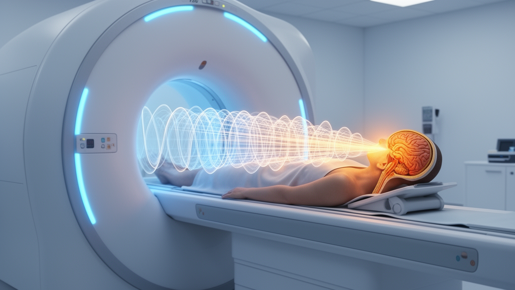
Magnetic resonance-guided focused ultrasound, or MRgFUS, has quietly become one of the most consequential shifts in modern neurosurgery. The technique burns a millimetric lesion deep inside the living brain without a single incision, using hundreds of intersecting ultrasound beams that converge on a target while an MRI scanner watches the temperature rise in real time. Regulators and clinicians have already embraced it for medication-refractory essential tremor and tremor-dominant Parkinson’s disease, where a unilateral thalamotomy of the ventral intermediate nucleus, the Vim, can quiet a shaking hand within hours. But as the technology spreads from specialized centers toward broader clinical adoption, a new problem has come into focus: the success of incisionless brain surgery now depends less on the ultrasound hardware and more on the precision of the neuroimaging that plans, guides, and evaluates every procedure.
That transition is the subject of a new editorial published in Frontiers in Radiology by Federico Bruno of San Salvatore Hospital in L’Aquila, Italy, and Sotirios Bisdas of University College London. Writing on 15 September 2026, the two neuroradiologists argue that imaging is no longer merely the navigation platform for sonication. Instead, they describe it as the connective tissue of the entire therapeutic pathway, stretching from target definition and intraoperative decision-making to mechanistic interpretation, outcome assessment, and potentially individualized prognosis. Their editorial frames a five-study Research Topic that spans direct thalamic visualization, multimodal localization of emerging circuit-based targets, longitudinal network analysis, radiomics-based outcome modeling, and quantitative assessment of staged bilateral treatment. Together, the studies position precision neuroimaging not as a single sequence or clever technique, but as an operational framework linking patient-specific anatomy to circuit organization, procedural execution, tissue response, and clinical follow-up.
The first contribution tackles a limitation that has shadowed Vim thalamotomy since its inception: reliance on indirect, atlas-based coordinates despite meaningful variation between individual brains. Aibar-Duran and colleagues used 3-Tesla white-matter-nulled MPRAGE imaging, a structural MRI sequence whose contrast nulls the signal of white matter to make gray-matter structures stand out, to directly visualize the Vim and its neighboring thalamic structures. They paired this direct anatomical information with a dual-lesion strategy designed to conform to the nucleus’s oblique anterior-superior orientation, an oblique axis that a single spherical ablation may not adequately cover. Their experience supports the feasibility of integrating direct anatomical visualization into routine MRgFUS planning and illustrates how target coverage can be adapted to individual anatomy. The authors are careful about scope: the report does not establish superiority over conventional targeting or provide comparative clinical validation. Its value is both practical and agenda-setting, shifting the question from where an atlas predicts the target to how confidently that target and its boundaries can be identified in the specific patient being treated.
Precision, as the editorial emphasizes, also requires honesty about what current imaging cannot yet resolve. Rivera and colleagues examined the pallidothalamic region, an emerging circuit-based target for Parkinson’s disease in which the relevant fiber pathways remain difficult to visualize directly even with state-of-the-art scanners. Their mini review integrates high-resolution structural MRI, inversion-recovery and susceptibility-based contrasts, and diffusion tractography, the technique that reconstructs fiber pathways from the directional diffusion of water molecules. Crucially, they highlight the mammillothalamic tract as a comparatively consistent adjacent landmark. The mammillothalamic tract is not proposed as the therapeutic target or as a fixed millimetric offset from it; rather, it may help orient the pallidothalamic corridor while simultaneously flagging a memory-related pathway that should be preserved. This distinction exemplifies a mature use of multimodal imaging: not to claim visibility that is not yet achievable, but to reduce anatomical uncertainty through complementary information. Prospective studies must still determine whether the approach improves targeting accuracy, safety, or clinical outcomes.
Once a lesion has been placed, imaging can interrogate consequences that extend far beyond the ablation site. Jiang and colleagues studied resting-state functional connectivity, measured with functional MRI while patients simply lay still, before and one year after unilateral Vim thalamotomy in 15 patients with essential tremor. Sustained improvement in the treated hand was accompanied by altered connectivity involving the Vim, the primary motor cortex, the dentate nucleus of the cerebellum, and distributed motor, sensory, attentional, visual, and visuospatial regions. Notably, changes in left Vim connectivity were associated with changes in tremor severity. These observations support a network-level interpretation of MRgFUS, in which a focal lesion is associated with remote and bilateral functional reorganization across the motor system and beyond. The authors and the editorial are equally clear about the limits: the findings do not establish causality or a validated predictive biomarker. Larger controlled cohorts, complemented by tractography and standardized cognitive assessment, will be needed to determine which connectivity changes mediate benefit, reflect compensation, or simply accompany recovery.
Where functional MRI looks outward from the lesion to the network, radiomics looks inward to quantify tissue patterns that conventional visual inspection may not capture. Innocenzi and colleagues analyzed 24-hour post-treatment T2-weighted MRI in 120 patients undergoing unilateral MRgFUS thalamotomy and trained a machine-learning model to classify clinically relevant tremor recurrence at 12 months. Recurrence occurred in 23 patients, roughly 19 percent of the cohort, and the internally cross-validated model achieved a balanced accuracy of 0.720 in the full cohort, with stronger performance in Parkinson’s disease than in essential tremor. Texture features, which capture the statistical regularities of lesion appearance at the pixel level, contributed most strongly to the model, suggesting that the early lesion phenotype may contain information related to treatment durability. The study is appropriately viewed as a pilot: its single-center design, modest event count, diagnostic subgroup differences, class imbalance, manual segmentation, and absence of external validation preclude immediate clinical deployment. Nevertheless, it offers a concrete route from routine post-procedural MRI toward testable, quantitative prognostic tools.
Paio and colleagues extend the precision-imaging agenda to staged bilateral thalamotomy, where reproducibility must be balanced against the risks of bilateral intervention. In their single-center cohort of 15 patients, most anatomical, procedural, and sonication parameters were comparable between first- and second-side procedures, as were lesion volumes at 24 hours and at approximately one month. Second-side targeting incorporated a small anterior-dorsal refinement rather than strict mirror symmetry, and adverse events were predominantly mild and transient. Intriguingly, neither lesion volume nor inter-side targeting displacement distinguished patients with gait disturbance persisting at one month, while individual imaging patterns suggested that lesion geometry, extension into adjacent structures, and network effects may all matter. The study thus makes an important conceptual point: in bilateral MRgFUS, precision is not synonymous with geometric symmetry, but with cautious, imaging-informed adaptation.
Read together, the five contributions form a coherent translational arc. Before treatment, structural and diffusion imaging can reduce uncertainty in target localization and identify structures at risk. During treatment, MRI provides thermometric feedback and supports clinically guided refinement of the ablation. After treatment, lesion volumetry, radiomics, and functional connectivity can characterize tissue response, procedural consistency, and distributed network change. This continuum matters because MRgFUS outcomes cannot be reduced to a coordinate, a peak temperature, or a lesion volume in isolation. Durable benefit and acceptable risk are more likely to depend on the interaction among patient anatomy, circuit architecture, lesion geometry, biological response, and functional reserve.
The next phase of the field, Bruno and Bisdas argue, should be defined by validation and integration. Harmonized acquisition, segmentation, tractography, and outcome definitions are needed to make results comparable across institutions. Prospective multicenter studies should combine structural, diffusion, functional, and quantitative lesion imaging with clinical and procedural variables, while testing models on genuinely external data. Patient-specific connectomics, computational modeling, radiomics, and explainable artificial intelligence could ultimately help refine target selection, anticipate benefit and adverse effects, and tailor follow-up, but their clinical value will depend on transparent uncertainty, reproducibility, and evidence that imaging-informed decisions actually improve outcomes. The editorial does not present a finished solution; more usefully, it defines a credible and testable roadmap. As MRgFUS moves toward more complex targets, staged bilateral strategies, and more individualized care, precision neuroimaging will be central to translating technical capability into durable, safe, and generalizable clinical benefit.
Subject of Research: Editorial: Precision neuroimaging for MRgFUS in neurological disorders
Article Title: Editorial: Precision neuroimaging for MRgFUS in neurological disorders
Article References: Bruno, F., & Bisdas, S. (2026). Editorial: Precision neuroimaging for MRgFUS in neurological disorders. Frontiers in Radiology, 6, Article 1979646. https://doi.org/10.3389/fradi.2026.1979646
Image Credits: AI Generated
DOI: 10.3389/fradi.2026.1979646
Keywords: Editorial, Precision, neuroimaging, MRgFUS, neurological, disorders, scientific research
Cite Scienmag News
Cassandra Pierce. (October 17, 2026). Incisionless Brain Surgery Meets Its Match: The Imaging Revolution Behind Focused Ultrasound. Scienmag. https://scienmag.com/incisionless-brain-surgery-meets-its-match-the-imaging-revolution-behind-focused-ultrasound/
Cassandra Pierce. "Incisionless Brain Surgery Meets Its Match: The Imaging Revolution Behind Focused Ultrasound." Scienmag, 17 October 2026, https://scienmag.com/incisionless-brain-surgery-meets-its-match-the-imaging-revolution-behind-focused-ultrasound/. Accessed 17 October 2026.
Cassandra Pierce. "Incisionless Brain Surgery Meets Its Match: The Imaging Revolution Behind Focused Ultrasound." Scienmag. October 17, 2026. https://scienmag.com/incisionless-brain-surgery-meets-its-match-the-imaging-revolution-behind-focused-ultrasound/


