


Purines are among the most consequential small molecules in biology. The adenine derivatives ATP, ADP, AMP, adenosine and adenine sit at the heart of energy transfer, RNA and DNA synthesis, and cellular signaling, while the guanine derivatives GTP, GDP, GMP and guanosine power protein synthesis and regulate signaling cascades. Defects in nearly every branch of purine metabolism are now recognized causes of human disease, from gout and kidney stones to devastating neurological disorders such as Lesch-Nyhan disease. Yet measuring the full purine landscape inside cells and tissues has long demanded slow, laborious chromatographic methods. A team at Emory University School of Medicine has now unveiled a faster, more sensitive technique that resolves and quantifies 15 of the most abundant biologically relevant purine metabolites in a single 33-minute run, roughly half the time of the method it replaces.
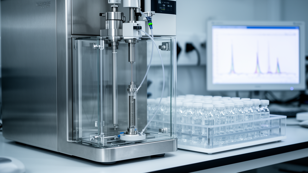
The new approach, described in Frontiers in Analytical Science by Rong Fu, Diane J. Sutcliffe, Ashok R. Dinasarapu, Ellen J. Hess and H. A. Jinnah, relies on ultra performance liquid chromatography coupled with photodiode array ultraviolet detection, commonly abbreviated UPLC-PDA. Unlike mass spectrometry-based metabolomics, which can be expensive and technically demanding, PDA detection exploits the strong ultraviolet absorbance of purines at 254 nanometers, allowing straightforward identification by retention time and spectral profile. The system consists of a Waters ACQUITY H-Class plus core with a quaternary solvent manager and an eLambda PDA detector, operating on a 2.1 by 100 millimeter reverse-phase C18 column packed with 1.8 micrometer particles at 40 degrees Celsius.
Separation chemistry is the crux of the method. Purines carry ionizable groups whose charge states shift dramatically with pH, so the Emory team designed a stepped gradient between two carefully formulated buffers. Buffer A contains 10 millimolar ammonium acetate and 1.5 millimolar tetrabutylammonium phosphate at pH 5.0, while buffer B contains 10 millimolar ammonium phosphate with the same ion-pairing reagent and 25 percent acetonitrile at pH 7.0. The tetrabutylammonium phosphate acts as an ion-pairing agent, taming the highly polar nucleotides so they interact with the reverse-phase column and elute as discrete peaks rather than co-eluting in the void volume. The gradient holds 100 percent buffer A for 11 minutes, ramps to 75 percent buffer B, then climbs to 100 percent buffer B before the column re-equilibrates for 9.5 minutes ahead of the next injection.
With this configuration, the method resolves all 15 target metabolites: ATP, ADP, AMP, adenosine, adenine, GTP, GDP, GMP, IMP, ZMP, guanosine, hypoxanthine, inosine, xanthine and uric acid. Only 5 microliters of sample are injected per run. Calibration with purified standards confirmed linear detection across a remarkable four-order-of-magnitude range from 0.1 to 100 micromolar, with all metabolites detectable down to approximately 100 nanomolar. Reproducibility proved equally impressive. When the same sample was analyzed five times within a single batch and across three batches on different days, the within-run coefficient of variance averaged 0.8 percent and the between-run variance averaged 1 percent for most analytes, comfortably under the 2 percent threshold the authors cite as evidence of excellent reliability.
The improvements over the team’s earlier HPLC-PDA method are substantial. The original technique required 65 minutes per sample; the new UPLC version cuts that to 33 minutes. Sensitivity is higher with half the sample volume, a critical advantage when working with scarce clinical specimens or small cell cultures. Perhaps most importantly, the new method cleanly discriminates GMP from IMP, two metabolites that co-eluted and could not be separately quantified with the older protocol. Because GMP and IMP occupy distinct positions in purine interconversion pathways, their separate measurement opens new analytical possibilities for studies of guanine nucleotide synthesis and purine salvage.
To demonstrate performance in genuine biological material, the researchers applied the method to induced pluripotent stem cells derived from three publicly available lines: KOLF2.0, NCRM1 and PGP-1. Each line was studied in matched pairs, with and without a c.508C>T stop codon mutation in the HPRT1 gene. This mutation abolishes hypoxanthine-guanine phosphoribosyltransferase, the salvage enzyme whose complete loss causes Lesch-Nyhan disease, a rare X-linked disorder marked by self-injurious behavior, dystonia and severe motor impairment. Cells were grown in six-well plates to roughly one million cells per well, with culture medium freshly replaced 24 hours before collection.
Sample preparation demanded meticulous attention to chemistry. For intracellular purines, cells were released by trypsinization, pelleted, and extracted in 0.2 molar perchloric acid, which instantly denatures the enzymes that would otherwise degrade labile nucleotides. Sonication at 4 degrees Celsius liberated the contents, and after centrifugation the acidic supernatant was neutralized with potassium carbonate, precipitating potassium perchlorate before final filtration through PVDF microcentrifuge tubes. For extracellular purines, 450 microliters of conditioned medium were acidified with perchloric acid to 0.1 molar and processed identically. Intracellular results were normalized to total cellular protein measured with a BCA assay, while extracellular results were corrected for culture volume and duration.
The biological findings were striking. In keeping with decades of prior work, the adenine derivatives dominated the intracellular purine pool, with ATP present in the greatest amounts, roughly 33 to 46 nanomoles per milligram protein across the lines, followed by GTP at 11 to 16 nanomoles per milligram. The adenylate energy charge, calculated as ([ATP] + 0.5 × [ADP]) / ([ATP] + [ADP] + [AMP]), ranged from 0.81 to 0.88, confirming that all cultures retained a healthy nucleotide energy state above the 0.80 benchmark typical of viable cells. Multivariate analysis of variance revealed significant main effects for both mutation status and cell type, along with a significant interaction between the two variables.
Most intracellular purines showed small or no changes in the HPRT1-mutant cells, but the extracellular compartment told a very different story. Conditioned medium contained no detectable purines except hypoxanthine, which was elevated by an average of 30-fold in the mutant lines, with individual increases ranging from 12-fold to as much as 225-fold depending on the genetic background. The explanation is elegant: without functional HPRT, the cells cannot recycle hypoxanthine back into nucleotides, so they simply dump it into the extracellular space. This finding underscores why measuring extracellular purines is essential, since changes outside the cell were far more obvious than those within it.
The authors acknowledge limitations. The method focuses on free purines and does not capture purines incorporated into DNA, RNA, NAD(H), NADP(H), FAD(H) or other complex molecules, though it can be adapted for some of these derivatives. Cyclic nucleotides and deoxynucleotides occur at levels too low for direct detection without prior sample concentration. The team also emphasizes the extraordinary speed of purine degradation: within seconds, ATP is dephosphorylated to ADP and then AMP, and within minutes AMP is further catabolized to adenosine, inosine, hypoxanthine and uric acid. Tissue measurements demand freeze-clamping in situ or focused microwave irradiation to halt degradation. Finally, the authors caution that isolated ATP measurements can be misleading, since they cannot distinguish energy impairment from wholesale purine depletion; only the simultaneous measurement of ATP, ADP and AMP, captured by the adenylate energy charge, provides a faithful picture of cellular energy state. With its speed, sensitivity and comprehensive coverage, the new UPLC-PDA method appears well suited to simultaneous analysis of the most abundant biologically relevant purines across blood, urine, cerebrospinal fluid and cultured cells, offering a powerful new lens on disorders of purine metabolism.
Subject of Research: Biomedical Analysis and Diagnostics
Article Title: Comprehensive measurement of purines in biological samples
Article References: Fu, R., Sutcliffe, D. J., Dinasarapu, A. R., Hess, E. J., & Jinnah, H. A. (2025). Comprehensive measurement of purines in biological samples. Frontiers in Analytical Science, 5, Article 1600781. https://doi.org/10.3389/frans.2025.1600781
Image Credits: AI Generated
DOI: 10.3389/frans.2025.1600781
Keywords: purine metabolism, UPLC-PDA, Lesch-Nyhan disease, hypoxanthine, adenylate energy charge, metabolomics, HPRT1, analytical chemistry, Comprehensive, measurement, purines, biological
Cite Scienmag News
Alexandra Wallace. (October 16, 2026). New UPLC Method Measures 15 Purine Metabolites in Half the Time. Scienmag. https://scienmag.com/new-uplc-method-measures-15-purine-metabolites-in-half-the-time/
Alexandra Wallace. "New UPLC Method Measures 15 Purine Metabolites in Half the Time." Scienmag, 16 October 2026, https://scienmag.com/new-uplc-method-measures-15-purine-metabolites-in-half-the-time/. Accessed 16 October 2026.
Alexandra Wallace. "New UPLC Method Measures 15 Purine Metabolites in Half the Time." Scienmag. October 16, 2026. https://scienmag.com/new-uplc-method-measures-15-purine-metabolites-in-half-the-time/


