


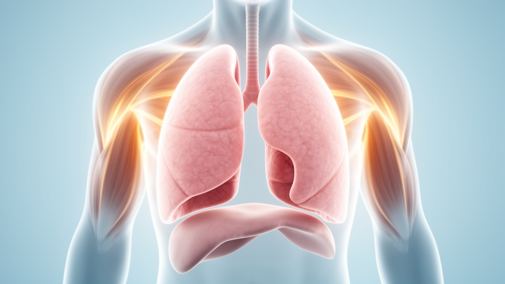
Shrinking lung syndrome, one of the rarest and most puzzling pulmonary complications of systemic lupus erythematosus, has long resisted systematic study. Now a comprehensive review published in Frontiers in Lupus has pooled data from 120 patients across 34 studies spanning nearly eight decades of medical literature, offering the clearest picture yet of how this enigmatic condition presents, how it can be diagnosed, and how it responds to treatment. The findings underscore both the distinctive clinical fingerprint of the syndrome and the persistent gaps in understanding its underlying biology.
Systemic lupus erythematosus is a chronic autoimmune disease in which the immune system attacks multiple organ systems, affecting an estimated 5.14 per 100,000 person-years worldwide. Women of childbearing age are disproportionately affected, being ten times more likely than men to develop the disease, and prevalence is highest among Black, Hispanic, and Asian populations. Pulmonary involvement occurs in a striking 20 to 90 percent of lupus patients, ranging from pleuritis and pleural effusion to acute pneumonitis, interstitial lung disease, and pulmonary hypertension. Against this backdrop, shrinking lung syndrome stands out as an uncommon but clinically important entity, estimated to affect between 0.5 and 1.53 percent of all lupus patients.
First described in 1965 by Hoffbrand and Beck, shrinking lung syndrome is characterised by progressive breathlessness, pleuritic chest pain, reduced lung volumes, and elevated hemidiaphragms on chest imaging, all in the absence of identifiable parenchymal lung disease. The syndrome has also been reported in other connective tissue diseases, including Sjögren’s syndrome, scleroderma, and rheumatoid arthritis, though it remains most strongly associated with lupus. What has made the condition so difficult to study is its rarity and the absence of standardised diagnostic criteria, forcing clinicians to diagnose it largely by excluding pneumonia, pulmonary embolism, heart failure, interstitial lung disease, and other more common causes of restrictive lung physiology.
The researchers, led by Nandini Bindal of Queen Mary University of London and Debasish Pyne of the Barts Health Lupus UK Centre of Excellence, conducted a systematic search of MEDLINE and Embase databases covering publications from January 1946 through December 2025. They identified 107 records, of which 34 met their inclusion criteria: 19 case reports, nine case series, five case series with literature reviews, and one cross-sectional cohort study. From these, they pooled data on 120 patients. The demographic profile mirrored that of lupus itself, with 86 percent of patients being female and ages ranging from 12 to 67 years, including several paediatric cases. The mean age across studies was approximately 33 years.
Clinically, the syndrome announced itself most often through breathlessness. Dyspnoea was present in 98 percent of patients for whom symptom data were available, while pleuritic chest pain affected 81 percent. Orthopnoea, fever, fatigue, arthralgia, and cough were also described, though less consistently quantified. In 22 percent of cases, shrinking lung syndrome was the initial manifestation of lupus, meaning that for some patients, respiratory symptoms were the first clue that they had an autoimmune disease at all. Among those who developed the syndrome after an established lupus diagnosis, the median interval from lupus diagnosis to syndrome onset was 48 months, with a remarkable range extending from concurrent onset to 38 years later. Symptom onset itself varied widely, from gradual progression over months to years to abrupt deterioration, including one case triggered by prednisolone withdrawal after two months of medication non-compliance.
Imaging findings reinforced the syndrome’s characteristic signature. Chest radiographs were performed in 88 percent of patients and computed tomography in 68 percent. Elevated hemidiaphragms were identified in 78 percent of the pooled cohort, reduced lung volumes in 43 percent, and basal or laminar atelectasis in 34 percent. Pleural effusions or pleural thickening appeared in 31 percent of patients. Importantly, lung parenchyma was generally preserved, with no evidence of significant fibrosis or diffuse interstitial changes, distinguishing the syndrome from interstitial lung disease. Some patients had entirely normal baseline imaging, with serial chest radiographs later revealing progressive diaphragmatic elevation, highlighting the dynamic nature of the condition. Diaphragmatic fluoroscopy and ultrasound, performed in a subset of patients, confirmed reduced diaphragmatic excursion in 19 patients and complete absence of movement in one.
Pulmonary function testing provided perhaps the most diagnostically useful pattern. A restrictive ventilatory defect was present in 91 percent of the 117 patients with available data. Forced vital capacity values ranged from 15 to 99 percent of predicted, with most patients showing moderate to severe restriction, and total lung capacity was similarly reduced. Yet the diffusing capacity for carbon monoxide, while reduced in the majority, was preserved or elevated in the transfer coefficient (KCO) in 90 percent of the 33 patients in whom it was measured. This disproportionate reduction in lung volumes relative to preserved gas transfer efficiency points to an extrapulmonary cause of restriction, offering clinicians a valuable clue that separates shrinking lung syndrome from interstitial lung disease, where both lung volumes and gas transfer are typically reduced together.
The pathophysiology of the syndrome remains contested, and the pooled data reflect this uncertainty. Phrenic nerve conduction studies, available in only four patients, yielded heterogeneous results: one patient had normal nerve potentials, another showed normal diaphragm twitch pressures on magnetic stimulation, and a third displayed absent electrical activity on the left hemidiaphragm with delayed responses on the right, consistent with a demyelinating phrenic neuropathy that improved after corticosteroid treatment. One patient underwent dynamic magnetic resonance imaging, which revealed normal movement of both diaphragms and all accessory respiratory muscles during forced ventilation, suggesting that diaphragmatic dysfunction may not always explain the restriction. Muscle biopsy findings from two other cases pointed instead to a myopathic mechanism, with one showing an endomysial T-lymphocytic infiltrate and another describing a CD20-positive B-cell-predominant myositis responsive to mycophenolate mofetil. These scattered observations suggest that shrinking lung syndrome may not be a single entity but rather the convergent clinical expression of several distinct immune-mediated mechanisms.
Treatment in the pooled cohort was dominated by corticosteroids, given to 93 percent of patients, typically as oral prednisolone at doses of 15 to 60 milligrams per day or as intravenous methylprednisolone pulses. Conventional immunosuppressants were frequently added, including azathioprine in 28 percent, cyclophosphamide in 19 percent, mycophenolate mofetil in 18 percent, and methotrexate in 8 percent. Rituximab, a B-cell-depleting monoclonal antibody, was used in 10 percent of patients, and belimumab in 2 percent, generally in refractory cases. Theophylline appeared in 10 percent of treatment regimens, though one patient developed paroxysmal supraventricular tachycardia and atrial flutter that persisted after discontinuation, underscoring the cardiac risks of this adjunct. Supportive measures such as inhaled beta-2 agonists and pulmonary physiotherapy were occasionally employed, and a structured inspiratory muscle training programme has shown promise in improving respiratory muscle strength and diaphragm thickness on ultrasound.
Outcomes were broadly favourable. Among 104 patients with reported symptomatic outcomes, 77 percent improved, 17 percent remained stable, and only 6 percent showed no improvement or worsened. Follow-up pulmonary function data in 85 patients demonstrated partial recovery in 64 percent, stabilisation of the restrictive pattern in 22 percent, and complete normalisation in just 7 percent. Notably, patients often reported symptomatic relief before measurable improvements in lung function, reinforcing the idea that the restriction reflects a reversible, functional process rather than fixed structural damage. Follow-up periods ranged from three weeks to 20 years, and one death was attributed to the syndrome across the entire cohort. The authors caution that the evidence base remains limited by its reliance on case reports and small series, with inconsistent reporting of investigations, treatments, and long-term follow-up. Nevertheless, the review makes a compelling case that early recognition of shrinking lung syndrome, combined with tailored immunosuppressive therapy, can achieve good outcomes for most patients, and that greater clinical awareness is essential to reduce the diagnostic delays that have historically plagued this rare but treatable complication of lupus.
Subject of Research: Shrinking lung syndrome as a rare pulmonary manifestation of systemic lupus erythematosus
Article Title: Shrinking lung syndrome in systemic lupus erythematosus: a systematic review
Article References: Bindal, N., & Pyne, D. (2026). Shrinking lung syndrome in systemic lupus erythematosus: a systematic review. Frontiers in Lupus, 4, Article 1845359. https://doi.org/10.3389/flupu.2026.1845359
Image Credits: AI Generated
DOI: 10.3389/flupu.2026.1845359
Keywords: shrinking lung syndrome, systemic lupus erythematosus, systematic review, diaphragmatic dysfunction, restrictive lung disease, corticosteroids, rituximab, pulmonary function tests, autoimmune disease, dyspnoea, immunosuppression, rare disease
Cite Scienmag News
Ophelia Keating. (October 16, 2026). Shrinking Lung Syndrome in Lupus: A Systematic Review of 120 Cases Reveals Distinctive Patterns. Scienmag. https://scienmag.com/shrinking-lung-syndrome-in-lupus-a-systematic-review-of-120-cases-reveals-distinctive-patterns/
Ophelia Keating. "Shrinking Lung Syndrome in Lupus: A Systematic Review of 120 Cases Reveals Distinctive Patterns." Scienmag, 16 October 2026, https://scienmag.com/shrinking-lung-syndrome-in-lupus-a-systematic-review-of-120-cases-reveals-distinctive-patterns/. Accessed 16 October 2026.
Ophelia Keating. "Shrinking Lung Syndrome in Lupus: A Systematic Review of 120 Cases Reveals Distinctive Patterns." Scienmag. October 16, 2026. https://scienmag.com/shrinking-lung-syndrome-in-lupus-a-systematic-review-of-120-cases-reveals-distinctive-patterns/


