


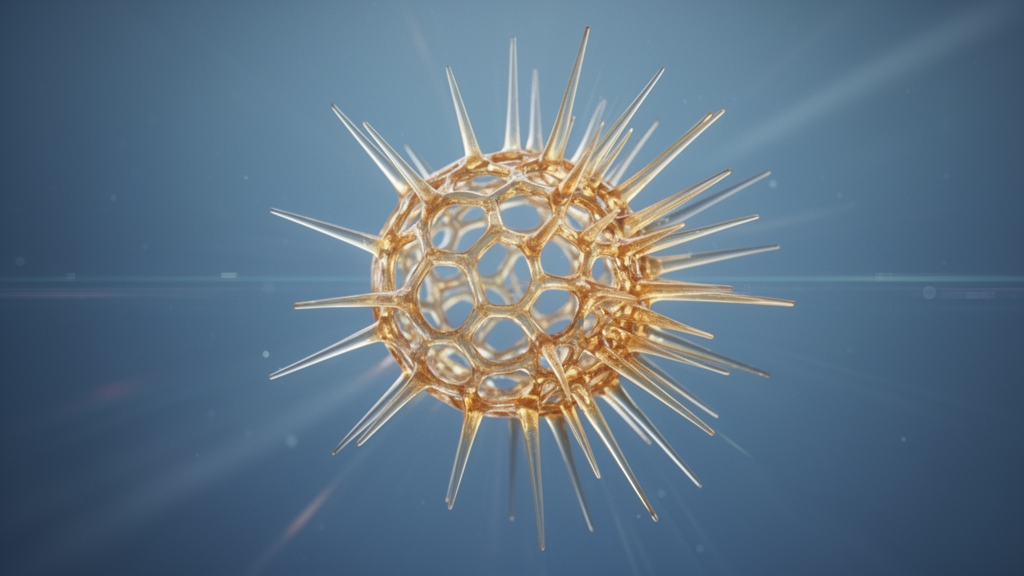
Beneath the surface of a limestone sample collected from western Newfoundland, a microscopic skeleton has been waiting nearly 480 million years to tell its story. That skeleton belonged to Protoentactinia gracilispinosa, a radiolarian — a single-celled marine organism that secretes an intricate test of amorphous silica — that lived during the latest Cambrian and earliest Ordovician periods. Using X-ray micro-computed tomography, researchers have now peered inside this delicate fossil in three dimensions and, for the first time, reconstructed the complete sequence of branching stages by which its skeleton was built. The findings, published in the journal Fossil Record, confirm long-standing assumptions about how this species grew, but they also reveal something unexpected: the skeleton did not develop symmetrically, as its subspherical final form might suggest, but instead grew with a pronounced directional bias.
Radiolarians are planktic Rhizaria, a diverse group of marine organisms whose siliceous skeletons make them among the most important microfossils for understanding early ocean ecosystems. Molecular clock studies suggest that radiolarians lacking skeletons may have existed since the Late Neoproterozoic, but the fossil record of polycystine radiolarians — those bearing skeletons — is only confirmed from the Cambrian Series 2, Stage 4. The Cambrian and Ordovician periods are therefore of key significance for the early evolutionary history of these heterotrophic plankton, and the siliceous skeletons of early Palaeozoic radiolarians testify to significant diversifications and evolutionary changes during this interval.
For the past 35 years, progress in understanding the taxonomy and biostratigraphy of early Palaeozoic radiolarians has relied largely on the discovery of well-preserved faunas and their description using two-dimensional imaging techniques such as scanning electron microscopy. While SEM has been invaluable, it cannot capture the full three-dimensional complexity of a radiolarian skeleton, particularly its most internal parts, which carry the greatest taxonomic significance. The recent application of micro-computed tomography to radiolarians has changed this, allowing researchers to observe the most internal architectures of these fossils in a non-destructive way and opening new avenues for understanding their internal organisation.
The species at the centre of the new study was first introduced by Kozur, Mostler and Repetski in 1996, based on Tremadocian (Lower Ordovician) material from Nevada. It was described as the type species of the newly-defined genus Protoentactinia and type-genus of the family Protoentactiniidae. The original description characterised it as bearing a spicular system initiated from a bar-centred entactinarian spicule with three terminal rays at each end, with the subglobular skeleton formed of a repeatedly branching pattern of spicules that accommodated a nearly spherical inner empty space. The species was later reported from upper Cambrian and lowermost Ordovician strata of western Newfoundland.
The single specimen analysed in the new study came from a basal Tremadocian radiolarian assemblage extracted by Lauren Pouille of the University of Lille during her PhD work, after hydrochloric acid leaching from a limestone sample of the Green Point section of the Cow Head Group in western Newfoundland, Canada. Protoentactinia gracilispinosa is abundant in this assemblage, representing approximately 15 percent of over 2,000 identified specimens, and has been well documented by Pouille and colleagues. Skeleton analysis was conducted at Ghent University’s Centre of X-ray Tomography in Belgium using a custom-built Nanowood micro-CT scanner operating at 70 kV and 4 W, with a voxel size of 900 nanometres. A total of 2,322 projections were acquired at an exposure time of 1,000 milliseconds per projection, resulting in a total scan time of around 40 minutes. The projections were reconstructed using Octopus Reconstruction software, during which ring filtering and beam hardening corrections were applied.
Stacks of two-dimensional images were then processed using the open-source 3D Slicer software, which is commonly used to extract structural models from images primarily in clinical and biomedical applications. Image segmentation delineates regions within an image — in this case, structures of the siliceous skeleton — and is essential for visualising and quantifying them. The segmentation was performed semi-automatically: by manually scanning all the slices in three dimensions and using the segmentation tools provided by 3D Slicer, the researchers delineated areas of interest by selecting corresponding grey levels. Each part or segment could then be coloured, facilitating the interpretation of each branch within the three-dimensional volume. Remarkably, although the physical specimen was inadvertently lost during transport after all imaging had been completed, the description presented in the study is based on the comprehensive microscopic documentation acquired prior to its loss.
The colouring dissection allowed the researchers to recognise eight successive stages of spicular branching that result in the formation of a subspherical spicular test displaying a practically empty inner space. The first stage is represented by the initial bar-centred entactinarian spicule, which displays two terminal trifurcating branchings developed at the two extremities of the median bar. The vast majority of branchings throughout the skeleton’s development are of the trifurcating type, though rare single-rayed, bifurcating, quadrifurcating, pentafurcating and hexafurcating branchings were also observed, with only one single case of a heptafurcating branching recorded.
The numbers tell a striking story of growth dynamics. The total number of new branchings and rays increases rapidly up to stage 5, after which both decrease. Stage 2 produced 6 new branchings and 18 new rays; stage 3 generated 17 branchings and 50 rays; stage 4 yielded 35 branchings and 108 rays; and stage 5 recorded the maximum, with 36 branchings and 109 rays. However, after stage 2, only a fraction of existing rays leads to the development of new branchings. Out of the 18 new rays developed at stage 2, only 17 produce new branchings at stage 3. Out of the 50 new rays formed during stage 3, only 35 produce new branchings during stage 4. More remarkably, only a third of the rays newly formed during stage 4 branch out during stage 5. Therefore, the dynamic of skeletal growth decreases after stage 4, even though stage 5 records the maximum number of rays ever produced for this specimen.
In terms of spatial development, nearly half of the final subspherical spicular test is formed by the end of stage 3. Starting with stage 4, shell growth becomes clearly unidirectionally and asymmetrically orientated, away from the initial spicule, establishing a clear heteropolarity in the skeleton growth and structural organisation while simultaneously forming a more complete subspherical test. The distally orientated development of the shell is more obvious during stage 5. Most of the branches developed during stage 6 are generated on the opposite side of the initial spicule and in such a way as to form a subspherical shell. The few new branchings and rays generated during stages 7 and 8 are also developed so as to increase the subspherical aspect of the test.
The discovery of heteropolar development is particularly significant. Until now, heteropolar development of a polycystine radiolarian skeleton had been recognised only for radiolarians of the order Nassellaria. The new study documents for the first time the heteropolar growth of the test in a primitive entactinarian species. The skeleton growth of Protoentactinia gracilispinosa recalls the heteropolar and unidirectional skeleton development described in Nassellaria, but without the axial symmetry characteristic of nassellarians, as the end product is a subspherical form. During the first two stages of skeletal growth, the type of branchings is always trifurcating, and since this type represents the vast majority of total branchings formed during skeleton formation, it may very well be that the trifurcating type is the standard, with all other types of branching representing possible malformations or simply part of the infraspecific variability of the species. However, additional material needs to be analysed in the future to assess this, as three specimens of the species illustrated recently in a PhD thesis remain pending analysis.
Few studies provide insights into the function of polycystine radiolarian skeletal morphology. The researchers suggest that the siliceous skeleton might have served as an anchoring structure for a better function of the axoflagellates and cytoplasmic extensions. They also speculate that Protoentactinia gracilispinosa had a cell structure similar to the periaxoplastitid-type, described from cellular observations of living radiolarians from the western Mediterranean. If such was indeed the case, the axoplast of the organism was likely positioned close to the initial spicule, and axoflagellates were all orientated antapically, away from the axoplast. Such an axoflagellate organisation would be consistent with the unidirectional and heteropolar skeletal growth observed, in spite of the final subspherical form.
The analysis confirms the initial description provided by Kozur and colleagues in 1996: the subglobular spongy test of this entactinarian species is formed by repeated branchings of rays starting from the two trifurcating branchings of the initial spicule formed out of each termination of the median bar. The skeleton is formed subsequently through seven additional stages of reiterated branchings, but the subglobular form of the test is essentially developed during stages 3, 4 and 5. The maximum number of branchings is manifested in stage 5, and the total number of new rays also reaches its maximum at stage 5, after which it decreases during stages 6, 7 and 8. Unidirectional and heteropolar skeletal growth, similar to that observed in nassellarians, is established here for the first time using micro-CT analysis. Heteropolarity is expressed by the directional asymmetry in the skeleton growth and architecture, starting with the very eccentric position of the initial spicule at the edge of an empty spherical space. The study highlights how modern imaging techniques can extract detailed biological information from microscopic fossils, even when the physical specimen itself is no longer available, and underscores the value of micro-CT for the taxonomic analysis of early Palaeozoic radiolarians.
Subject of Research: X-ray tomography reveals the skeletal growth pattern of the early radiolarian Protoentactinia gracilispinosa
Article Title: Skeletal growth pattern of Protoentactinia gracilispinosa Kozur, Mostler & Repetski, 1996, a late Cambrian – Early Ordovician radiolarian species, revealed by X-ray tomography
Article References: Harutyunyan, L., Habert, R., Schröer, L., Cnudde, V., & Danelian, T. (2026). Skeletal growth pattern of Protoentactinia gracilispinosa Kozur, Mostler & Repetski, 1996, a late Cambrian – Early Ordovician radiolarian species, revealed by X-ray tomography. Fossil Record, 29(2), 759-764. https://doi.org/10.3897/fr.29.200748
Image Credits: AI Generated
DOI: 10.3897/fr.29.200748
Keywords: radiolaria, micro-CT, palaeontology, Cambrian, Ordovician, entactinarian, skeletal growth, Newfoundland, Skeletal, growth, pattern, Protoentactinia
Cite Scienmag News
Gavin Prescott. (October 15, 2026). X-ray Tomography Reveals Directional Growth in an Early Radiolarian Skeleton. Scienmag. https://scienmag.com/x-ray-tomography-reveals-directional-growth-in-an-early-radiolarian-skeleton/
Gavin Prescott. "X-ray Tomography Reveals Directional Growth in an Early Radiolarian Skeleton." Scienmag, 15 October 2026, https://scienmag.com/x-ray-tomography-reveals-directional-growth-in-an-early-radiolarian-skeleton/. Accessed 15 October 2026.
Gavin Prescott. "X-ray Tomography Reveals Directional Growth in an Early Radiolarian Skeleton." Scienmag. October 15, 2026. https://scienmag.com/x-ray-tomography-reveals-directional-growth-in-an-early-radiolarian-skeleton/


