


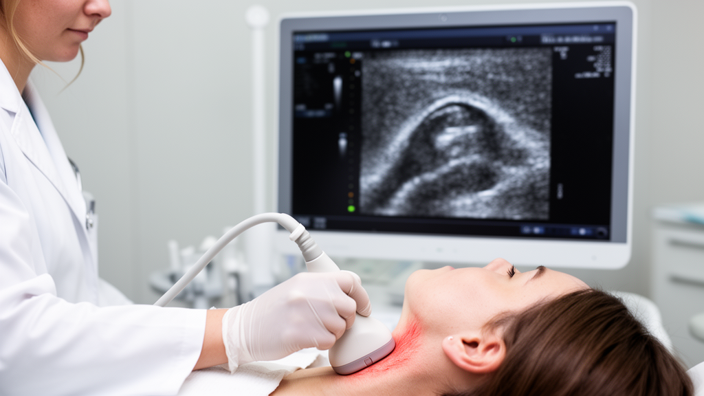
Every heartbeat sends a pulse of pressure down the carotid arteries, the twin vessels that carry blood to the brain, and in healthy people those artery walls flex and recoil thousands of times a day without complaint. In people with metabolic syndrome, however, that silent mechanical dance begins to falter long before any plaque or blockage appears. A new cross-sectional study published in BMC Medical Imaging suggests that a sophisticated ultrasound technique called speckle-tracking carotid strain ultrasonography, or STCS-US, can detect this hidden deterioration in remarkable detail, measuring not just how stiff an artery has become but how poorly it deforms with each pulse.
Metabolic syndrome is one of the most common and underappreciated health threats of modern life. Defined by the clustering of central obesity, insulin resistance, dyslipidemia, and hypertension, it affects roughly one quarter of the adult population worldwide. Each of these components independently raises cardiovascular risk, but together they act synergistically: the risk of heart disease and death climbs steadily as the number of metabolic abnormalities increases. What has frustrated clinicians for decades is that conventional imaging tools, such as carotid intima-media thickness measurements and plaque screening, capture only the structural aftermath of vascular disease. By the time arteries visibly thicken or develop plaques, the functional damage may have been accumulating for years.
The research team, led by investigators at Aydın Adnan Menderes University in Türkiye, set out to test whether STCS-US could fill that diagnostic gap. The technique borrows an idea from cardiac imaging: speckle-tracking software follows the natural acoustic patterns, or speckles, within tissue frame by frame, allowing researchers to quantify how the arterial wall moves, stretches, and recovers throughout the cardiac cycle. Rather than relying on a single stiffness number, the method yields a multidimensional portrait of arterial mechanics, encompassing both how rigid the vessel is and how much it actually deforms under pressure.
The study enrolled 100 participants, evenly split between 50 patients diagnosed with metabolic syndrome according to the National Cholesterol Education Program Adult Treatment Panel III criteria and 50 age- and sex-matched controls without the condition. The researchers retrieved archived carotid Doppler ultrasound cine-loop images from their institutional picture archiving system and analyzed them offline using the Arterial Analysis software on a Samsung V7 ultrasound system. From each participant, the software quantified 22 vascular parameters: five stiffness measures, including the beta-stiffness index, arterial compliance, arterial distensibility, elastic modulus, and pulse wave velocity, and six strain measures covering radial and circumferential displacement, strain, and strain rate, each captured in both longitudinal and transverse imaging planes.
The results were striking. Patients with metabolic syndrome showed significantly elevated stiffness indices, including higher elastic modulus and pulse wave velocity, alongside reduced compliance and distensibility compared with controls. But the most revealing findings came from the strain analysis. Both radial and circumferential strain percentages were significantly lower in the metabolic syndrome group, indicating that their artery walls simply could not stretch as far with each heartbeat. Intriguingly, these deformation abnormalities were most pronounced in the transverse imaging plane, where all stiffness parameters and both strain percentages differed between groups with p-values below 0.001, suggesting that the cross-sectional view of the artery may be particularly sensitive to early metabolic damage.
Perhaps the most clinically important observation was dose-dependent: the more metabolic syndrome components a participant had, the worse their arterial mechanics became. The number of diagnostic criteria correlated positively with stiffness indices such as the beta-stiffness index, elastic modulus, and pulse wave velocity, with the strongest associations appearing in the transverse plane. Conversely, compliance measures and strain percentages showed significant inverse correlations with metabolic burden. In other words, the technique did not merely separate patients with metabolic syndrome from healthy controls; it appeared to grade the cumulative damage inflicted by each additional risk factor, from elevated waist circumference to high triglycerides to raised fasting glucose.
To understand which factors truly drove these vascular changes, the team employed Classification and Regression Tree analysis, a machine-learning approach that ranks the relative importance of candidate variables. Metabolic syndrome status emerged as one of the most influential determinants across multiple parameters, topping the models for transverse-plane beta-stiffness index and elastic modulus. Body mass index carried the greatest weight for transverse-plane pulse wave velocity, while age dominated the models for radial and circumferential strain. Notably, conventional structural markers such as carotid intima-media thickness and plaque presence, along with sex, generally scored low in importance, hinting that functional arterial mechanics may carry information that structural imaging misses.
The biological story behind these numbers is well established. Insulin resistance, oxidative stress, endothelial dysfunction, and chronic low-grade inflammation promote remodeling of the extracellular matrix within the arterial wall, replacing elastic fibers with stiffer collagen. Hypertension and atherogenic dyslipidemia accelerate this process further. The metabolic syndrome group in this study displayed the expected biochemical signature: higher triglycerides, lower HDL cholesterol, elevated uric acid, increased white cell counts, and raised alanine aminotransferase levels suggestive of fatty liver involvement, all of which have been independently linked to arterial stiffening in previous research. Interestingly, LDL cholesterol was actually lower in the metabolic syndrome group, a discrepancy the authors attribute to the higher prevalence of lipid-lowering therapy among those patients, reinforcing the idea that atherogenic dyslipidemia may be a better vascular risk marker in this population than LDL alone.
The authors are careful to frame STCS-US as a complement rather than a replacement for established techniques. Applanation tonometry-based pulse wave velocity remains the gold standard for assessing arterial stiffness and is a proven independent predictor of cardiovascular events, but it reflects global arterial rigidity and says nothing about dynamic wall deformation during the cardiac cycle. Strain parameters, by contrast, probe how the vessel wall actually behaves under load, and prior studies have suggested that reduced carotid strain may carry prognostic information beyond conventional stiffness measures, including predictive value for stroke and acute coronary syndrome in metabolic populations. The combined impairment in both domains observed here suggests that metabolic clustering exerts an early, broad functional impact on arterial mechanics that a single stiffness metric cannot fully capture.
Honest limitations temper the excitement. The retrospective, single-center design precludes causal inference, the study was not powered to assess clinical outcomes, and differences in medication exposure between groups may have introduced residual confounding, potentially blunting the observed differences. Only intra-observer reproducibility was evaluated, yielding good agreement with an intraclass correlation coefficient of 0.800 and a coefficient of variation of 7.8 percent, while inter-observer and image-acquisition reproducibility remain untested. The authors emphasize that their findings are hypothesis-generating and call for prospective, multicenter studies with standardized acquisition protocols to determine whether integrated mechanical assessment can genuinely improve risk stratification. Still, the prospect is tantalizing: a routine carotid ultrasound, already performed in millions of patients, could be upgraded with speckle-tracking software to reveal not just what arteries look like, but how they are failing, potentially identifying the people whose metabolic burden is quietly stiffening their blood vessels years before a heart attack or stroke makes it impossible to ignore.
Subject of Research: Speckle-tracking carotid strain ultrasonography for assessing vascular dysfunction in metabolic syndrome
Article Title: Speckle-tracking carotid strain ultrasonography for assessment of vascular dysfunction in metabolic syndrome: a cross-sectional study
Article References: Topan, E. D., Varol, A., Uysal, H. B., Ömürlü, İ. K., & Gök, M. (2026). Speckle-tracking carotid strain ultrasonography for assessment of vascular dysfunction in metabolic syndrome: a cross-sectional study. BMC Medical Imaging, 26(1), Article 445. https://doi.org/10.1186/s12880-026-02686-5
Image Credits: AI Generated
DOI: 10.1186/s12880-026-02686-5
Keywords: metabolic syndrome, arterial stiffness, speckle-tracking, carotid ultrasonography, vascular dysfunction, pulse wave velocity, arterial strain, subclinical atherosclerosis, cardiovascular risk, ultrasound imaging, insulin resistance, carotid artery
Cite Scienmag News
Ophelia Keating. (October 15, 2026). Ultrasound Speckle-Tracking Reveals Hidden Artery Damage in Metabolic Syndrome. Scienmag. https://scienmag.com/ultrasound-speckle-tracking-reveals-hidden-artery-damage-in-metabolic-syndrome/
Ophelia Keating. "Ultrasound Speckle-Tracking Reveals Hidden Artery Damage in Metabolic Syndrome." Scienmag, 15 October 2026, https://scienmag.com/ultrasound-speckle-tracking-reveals-hidden-artery-damage-in-metabolic-syndrome/. Accessed 15 October 2026.
Ophelia Keating. "Ultrasound Speckle-Tracking Reveals Hidden Artery Damage in Metabolic Syndrome." Scienmag. October 15, 2026. https://scienmag.com/ultrasound-speckle-tracking-reveals-hidden-artery-damage-in-metabolic-syndrome/


