


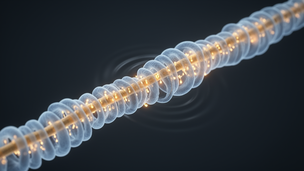
Every message that travels through the brain depends on speed, and speed depends on insulation. The fatty wrapping called myelin, laid down by specialized glial cells known as oligodendrocytes, allows electrical impulses to jump rapidly along nerve fibers instead of crawling through them. But myelin is not a uniform coating. In the central nervous system, the individual segments of myelin, each one a discrete sheath along an axon, vary enormously in length, differing by as much as an order of magnitude from one segment to the next. That variation is not random noise. It is now understood to be a fundamental design feature of neural circuitry, one that tunes the precise timing of signals arriving at their destinations and, by extension, shapes behavior itself. A new study published in PLOS Biology has finally identified a molecular mechanism that explains how these lengths are set, and the answer turns out to involve a remarkable piece of cellular engineering: a pressure-sensitive ion channel that lets oligodendrocytes literally feel the thickness of the nerve fibers they wrap.
The research, led by Amanda R. Young and colleagues in the laboratory of Marie E. Bechler, addresses a question that has lingered in neuroscience for decades. Since the earliest electron micrographs of nervous tissue, investigators have documented a striking correlation: the larger the diameter of an axon, the longer the myelin sheath that covers it. This scaling relationship holds across the central nervous system and is thought to be critical for synchronizing signal conduction. Longer internodes allow impulses to travel farther between the small gaps at nodes of Ranvier where the signal must be regenerated, which means that matching sheath length to axon caliber helps equalize conduction delays across fibers of different sizes. Without this coordination, circuits that depend on precisely timed inputs would fall out of rhythm. Yet despite decades of observation, no one knew how an oligodendrocyte, a cell that can extend dozens of separate myelin sheaths simultaneously, could measure the diameter of each underlying fiber and translate that measurement into a sheath of the appropriate length.
A crucial piece of groundwork came from the Bechler laboratory’s earlier work with a synthetic axon culture system. Rather than relying on living neurons, whose diameters and molecular compositions are difficult to control, the team grew rat oligodendrocytes on artificial fibers of defined width. The result was unambiguous: axon diameter alone was sufficient to instruct oligodendrocytes to build myelin sheaths of characteristic lengths. The glial cells did not need complex biochemical conversations with neurons to get the geometry right. The physical dimension of the fiber was enough. What remained unknown was the sensing mechanism, the molecular apparatus by which an oligodendrocyte converts the curvature or circumference of its substrate into an instruction to elongate its sheath to a particular extent.
The new study answers that question by pointing to Piezo1, a mechanosensitive ion channel that has become one of the most celebrated molecular sensors in modern cell biology. Piezo channels open in response to mechanical forces, including membrane tension, and convert physical stimuli into electrical and chemical signals within the cell. The researchers found that Piezo1 is positioned to serve exactly the role that diameter-sensing demands. When an oligodendrocyte begins wrapping a fiber, the geometry of the ensheathment differs depending on the caliber of that fiber, and those geometric differences can alter mechanical tension in the glial membrane. Piezo1, by transducing that tension into intracellular signaling, provides a plausible conduit from physical measurement to biological output.
Perhaps the most conceptually important finding of the study is that the regulation is local. A single oligodendrocyte can generate myelin sheaths of vastly different lengths on the same cell, depending on the diameters of the individual axons it contacts. This means the cell does not operate under a single global program that sets all of its sheaths to one length. Instead, each individual sheath behaves as an independent sensor-actuator unit, reading the diameter of the specific fiber beneath it and elongating accordingly. This local autonomy explains how one cell can simultaneously produce a short segment on a thin axon and a long segment on a thick one, a feat that would be impossible under centralized control. The implication is that myelin patterns across the brain emerge from countless independent diameter measurements, each performed at the level of a single sheath.
To confirm that the mechanism operates not just in a dish but in a living animal, the team turned to mouse models in which Piezo1 could be conditionally deleted from oligodendrocyte lineage cells. The in vivo results mirrored the in vitro findings with a striking specificity. Loss of Piezo1 impaired the elongation of myelin sheaths on large-diameter axons, precisely the fibers where the diameter-to-length scaling relationship is most demanding. Sheaths on smaller fibers were comparatively spared. This selective effect strongly supports the idea that Piezo1 is not a general-purpose growth factor for myelin but a dedicated component of the diameter-sensing machinery, engaged most heavily when the glial cell must accommodate a thick fiber.
Equally revealing is what the researchers did not find. Conditional loss of Piezo1 had no detectable impact on myelin thickness, the other major parameter of myelin architecture. This dissociation is significant because it demonstrates that the two defining dimensions of a myelin segment, its length along the axon and its thickness around it, are controlled by separable mechanisms. Myelin thickness has been the focus of most prior research on activity-dependent myelination and glial regulation, and many molecular pathways have been implicated in controlling how compact the wrapping becomes. The new work establishes that length is governed by its own rules, and that those rules involve mechanotransduction rather than the biochemical signaling pathways traditionally studied in the myelination field.
The authors propose that Piezo1 provides a mechanism for establishing what they describe as hard-wired myelin sheath patterns. In this view, the brain’s map of myelin segment lengths is not assembled gradually through experience-dependent refinement alone, but is substantially specified from the start by the intrinsic geometry of the axonal network. Oligodendrocytes, by transducing axon diameter through a mechanosensitive channel, generate myelin segments whose lengths are dictated by the physical structure of the circuit they invade. This does not rule out additional layers of regulation, including activity-dependent plasticity, but it establishes a baseline architecture that emerges from cellular mechanics. Such a hard-wired system would ensure that even before a circuit has been exercised, its conduction properties are already tuned to the dimensions of its component fibers.
The broader implications reach into several areas of neuroscience and medicine. Conduction timing is central to virtually every cognitive and motor function, and disruptions in myelin patterns are implicated in conditions ranging from multiple sclerosis to schizophrenia and autism spectrum disorders, where white matter abnormalities have been repeatedly documented. If sheath length is a critical variable in circuit timing, then understanding how it is set, and how it might be restored after injury or disease, becomes a therapeutic question in its own right. Current remyelination strategies focus largely on restoring the presence of myelin, with little attention to whether the regenerated segments adopt the correct lengths. A molecular target like Piezo1 suggests that future interventions might need to consider not just whether axons are remyelinated, but whether the new sheaths are geometrically appropriate for the fibers they cover.
The study also adds a compelling chapter to the growing recognition that mechanobiology shapes the nervous system in profound ways. Cells throughout the body use mechanical cues to make developmental decisions, and the finding that myelinating glia rely on a mechanosensitive channel to read the dimensions of their targets reinforces the idea that physical forces are not incidental to neural development but are actively encoded into its molecular machinery. An oligodendrocyte, in this picture, is not merely an insulating wrapper but a precision instrument, measuring the caliber of each fiber it encounters and fabricating a sheath whose length is calibrated to that measurement. The elegance of the solution, using a single ion channel to convert axon geometry into sheath architecture, exemplifies the kind of economical design that evolution so often produces, and it transforms our understanding of how the brain’s wiring achieves the timing precision on which every thought and movement depends.
Subject of Research: Mechanosensing by oligodendroglial Piezo1 in scaling central nervous system myelin sheath length to axon diameter
Article Title: Myelin sheath lengths in the central nervous system scale to axon diameter via oligodendroglial Piezo1
Article References: Myelin sheath lengths in the central nervous system scale to axon diameter via oligodendroglial Piezo1. (n.d.). https://doi.org/10.1371/journal.pbio.3003992
Image Credits: AI Generated
DOI: 10.1371/journal.pbio.3003992
Keywords: myelin, oligodendrocytes, Piezo1, axon diameter, mechanotransduction, central nervous system, sheath length, conduction timing, PLOS Biology, neuroscience, myelination, ion channels
Cite Scienmag News
Cassandra Pierce. (October 9, 2026). How Nerve Cells Measure Their Wires: Piezo1 Sets Myelin Length to Match Axon Size. Scienmag. https://scienmag.com/how-nerve-cells-measure-their-wires-piezo1-sets-myelin-length-to-match-axon-size/
Cassandra Pierce. "How Nerve Cells Measure Their Wires: Piezo1 Sets Myelin Length to Match Axon Size." Scienmag, 9 October 2026, https://scienmag.com/how-nerve-cells-measure-their-wires-piezo1-sets-myelin-length-to-match-axon-size/. Accessed 9 October 2026.
Cassandra Pierce. "How Nerve Cells Measure Their Wires: Piezo1 Sets Myelin Length to Match Axon Size." Scienmag. October 9, 2026. https://scienmag.com/how-nerve-cells-measure-their-wires-piezo1-sets-myelin-length-to-match-axon-size/


