


Researchers from NYU Langone Health are presenting a series of advances in corneal medicine and whole-eye transplantation at the American Academy of Ophthalmology annual meeting and the Cornea and Eye Banking Forum, held in New Orleans from October 9 to 12. The presentations span the spectrum of modern eye care: a landmark clinical trial showing that a simple topical medication can spare many patients a corneal transplant, a structured treatment framework for abnormal blood vessel growth in the cornea, the recognition of a vision-threatening nutritional deficiency that evades classic diagnosis, and a review of scientific progress toward total human eye transplantation. Kathryn A. Colby, the Elisabeth J. Cohen, MD, Professor of Ophthalmology and chair of the Department of Ophthalmology at NYU Grossman School of Medicine, framed the program as a reflection of the department’s mission, noting that the presentations advance a therapy that can spare patients a transplant, refine everyday clinical tools, and sharpen the diagnoses that save eyes and sometimes lives.
The most clinically consequential of the findings concerns Fuchs endothelial corneal dystrophy, a progressive genetic disease of the cornea’s inner cell layer that affects an estimated 4 to 7 percent of people in the United States and accounts for roughly one-third of the corneal transplants performed each year. The inner endothelium works as a fluid pump that keeps the cornea clear; when its cells progressively fail, the cornea swells, clouds, and vision deteriorates. For decades, the standard remedy for advanced disease has been replacement of the cornea with donor tissue, a strategy that works but carries the logistical, immunological, and lifelong follow-up burdens of transplant surgery.
A surgical alternative known as Descemet stripping only, or DSO, was pioneered in the United States by Dr. Colby. The procedure removes only the diseased central sheet of endothelial cells, leaving the patient’s healthier peripheral cells to migrate inward and repopulate the cornea. The approach eliminates the need for donor tissue in suitable patients, but recovery depends on how quickly and completely those remaining cells repopulate the central cornea. The new results address precisely that bottleneck, using a topical drug to accelerate the cellular healing process after surgery.
The phase 2 trial, known as Ripasudil (K-321-201), was the first multicenter, placebo-controlled randomized study of a Rho-associated protein kinase, or ROCK, inhibitor eyedrop administered after DSO. ROCK inhibitors are known to promote endothelial cell migration and proliferation and to discourage the cellular enlargement that marks endothelial failure. In the study, 65 patients at 38 sites across five countries were randomly assigned to receive the topical medication four times daily, twice daily, or a placebo for 12 weeks after undergoing DSO. Patients receiving the medicine achieved a central endothelial cell density of 531 cells per square millimeter, compared with 228 in the placebo group, indicating substantially better preservation and repopulation of the critical pumping layer.
The clinical outcomes were even more striking than the cell counts. Corneal swelling cleared completely in 17 of 21 patients using ripasudil, or 81 percent, compared with 2 of 22 patients, or 9 percent, in the placebo arm. Only 10 percent of patients in the medication group required a rescue corneal transplant, versus 27 percent of those on placebo. More than 95 percent of participants completed the one-year study, and no patient stopped treatment because of a side effect. The paper, co-authored by Dr. Colby, appeared as an American Ophthalmological Society thesis in the American Journal of Ophthalmology, and Dr. Colby presented the findings along with updates on the ongoing phase 3 trials during the Cornea and Eye Banking Forum. In her remarks, she emphasized that what matters most to patients is not a cell count but how quickly and predictably their vision returns and whether they can avoid a transplant altogether, adding that the trial provides high-quality evidence that a simple eye drop can do exactly that after DSO.
A second major theme at the meeting was corneal neovascularization, the abnormal growth of blood vessels into the normally clear cornea, a condition that affects an estimated 1.4 million people worldwide each year and is unilateral in about 76 percent of cases. It is most often driven by infectious keratitis, trauma, contact lens complications, and other ocular surface disease. Vascular invasion of the cornea not only degrades visual clarity directly but also dramatically raises the risk of rejection if the patient later needs a corneal transplant. Mohammad H. Dastjerdi, associate professor of ophthalmology at NYU Grossman School of Medicine, served as senior instructor for a course at the academy meeting outlining a staged treatment arsenal matched to the biology of the offending vessels.
The course’s central principle is that not all corneal vessels behave alike, and therapy must be matched to vessel maturity after the underlying trigger is treated. Newly formed, active vessels respond best to corticosteroids and anti-VEGF therapy such as bevacizumab, which suppresses the vascular growth signals sustaining them. Mature, lipid-laden vessels, which can be fully encased in protective pericytes, typically require destructive approaches such as fine-needle diathermy or mitomycin intravascular chemoembolization, abbreviated MICE. Faculty presented pooled data from an international retrospective series of 100 eyes treated with MICE between 2021 and 2024 after medical therapy failed. Surgeons reported full or partial vessel regression in approximately 81 percent of eyes at six to 12 months, and masked image analysis showed a mean 56 percent reduction in vessel area over the same interval. Best-corrected vision improved significantly, about 22 percent of eyes required repeat treatment, no infections occurred, and among 18 eyes that went on to corneal transplant, no rejections or graft failures were reported over at least one year of follow-up. Dr. Dastjerdi stressed that corneal neovascularization is not one disease and should not be met with one treatment, because the depth, caliber, and above all the age of the vessels dictate which tool to reach for, and combination therapy is often what finally breaks the cycle.
A third presentation sounded an urgent diagnostic alarm. At the Cornea and Eye Banking Forum, Dr. Dastjerdi described a retrospective case series of 17 adults with laboratory-confirmed vitamin A deficiency who developed corneal epithelial defects, ulceration, corneal melting, descemetocele, or perforation, yet none showed the classic outward signs clinicians are trained to recognize, such as conjunctival xerosis, Bitot’s spots, or clinically apparent corneal drying. The cohort included nine women and eight men. Nine patients, or 53 percent, presented with a descemetocele or frank corneal perforation, and every perforation occurred in a patient whose vitamin A level was below 10 micrograms per deciliter. Eight patients, or 47 percent, required tectonic keratoplasty, a structural corneal graft performed to preserve the integrity of the eye, and one additional patient died before planned surgery. Three patients, or 18 percent, died of systemic disease complications before their eye disease could be stabilized, and blood tests across the cohort revealed low levels of albumin, hemoglobin, magnesium, calcium, and vitamin D. Dr. Dastjerdi explained that these patients did not look like the textbook picture of vitamin A deficiency, which is precisely why the diagnosis is missed until the cornea has already melted, and he urged that unexplained corneal breakdown or ulceration alongside low albumin, anemia, or low magnesium should prompt a serum vitamin A level.
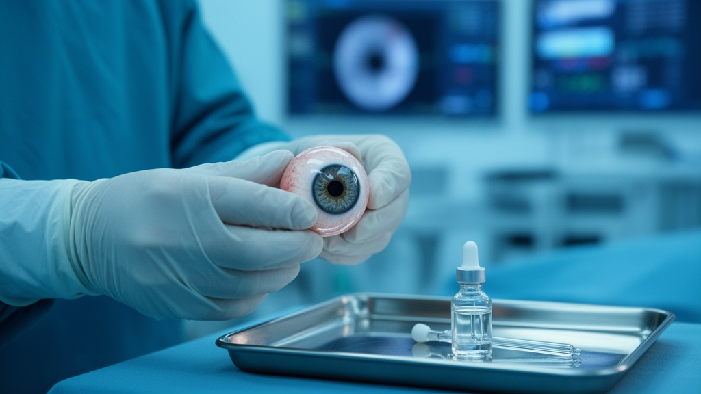
The program’s most ambitious offering looked beyond the cornea entirely. Vaidehi S. Dedania, associate professor of ophthalmology at NYU Grossman School of Medicine, participated in a symposium reviewing progress toward total human eye allotransplantation, or THEA, a frontier concept aimed at transforming treatment for irreversible blindness. Dr. Dedania was part of the NYU Langone team that completed the world’s first successful whole-eye and partial-face transplant in 2023. The obstacles to whole-eye transplantation are formidable: the complexity of orbital anatomy, and the intricacies and sensitivities of the optic nerve and retinal structures make functional restoration far harder than anatomical replacement. NYU Langone is part of a 56 million dollar effort through the federal Advanced Research Projects Agency for Health’s VISION Strategies for Whole Eye Transplant program, whose name captures the key research pillars of viability, imaging, surgical technique, immunomodulation, ocular preservation, and neuroregeneration. At the symposium, speakers discussed innovations in medical devices, novel surgical methods combined with advances in biostasis and perfusion, the newest successes in promoting long-distance optic nerve regeneration, and improvements in immunomodulation to prevent rejection.
Taken together, the New Orleans presentations illustrate a discipline in transition, in which the cornea is increasingly treated pharmacologically rather than replaced, vascular disease is managed by stratifying therapy to the biology of individual vessels, clinicians are being taught to detect a blinding nutritional disorder before it destroys tissue, and the first steps toward transplanting the entire human eye are being mapped through federally funded, multidisciplinary research. For patients with Fuchs dystrophy, the ripasudil results suggest that a future involving eyedrops rather than donor tissue is coming into view, while the broader message of the meeting is that careful diagnosis, staged therapy, and audacious long-horizon science can advance together within a single clinical enterprise.
Subject of Research: Corneal disease therapies and whole-eye transplant advances presented at the American Academy of Ophthalmology 2026 meeting
Article Title: NYU Langone Health ophthalmology faculty present advances in eye care at AAO 2026
Article References: NYU Langone Health ophthalmology faculty present advances in eye care at AAO 2026. (n.d.). Original publication
Image Credits: AI Generated
DOI: Not provided
Keywords: corneal transplant, Fuchs endothelial corneal dystrophy, Descemet stripping only, ripasudil, ROCK inhibitor, corneal neovascularization, anti-VEGF therapy, vitamin A deficiency, whole-eye transplant, optic nerve regeneration, ARPA-H, ophthalmology
Cite Scienmag News
Ophelia Keating. (October 9, 2026). Eye Drops, Hidden Deficiencies and Whole-Eye Transplants Take Center Stage at AAO 2026. Scienmag. https://scienmag.com/eye-drops-hidden-deficiencies-and-whole-eye-transplants-take-center-stage-at-aao-2026/
Ophelia Keating. "Eye Drops, Hidden Deficiencies and Whole-Eye Transplants Take Center Stage at AAO 2026." Scienmag, 9 October 2026, https://scienmag.com/eye-drops-hidden-deficiencies-and-whole-eye-transplants-take-center-stage-at-aao-2026/. Accessed 9 October 2026.
Ophelia Keating. "Eye Drops, Hidden Deficiencies and Whole-Eye Transplants Take Center Stage at AAO 2026." Scienmag. October 9, 2026. https://scienmag.com/eye-drops-hidden-deficiencies-and-whole-eye-transplants-take-center-stage-at-aao-2026/


