


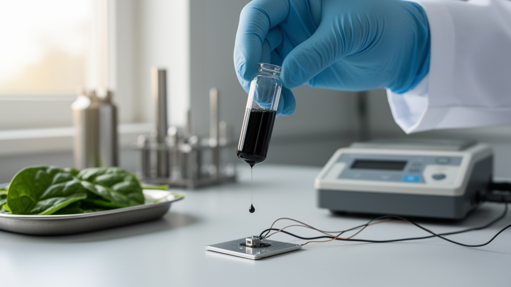
In a development that could reshape how clinicians monitor the brain’s most famous signaling molecule, researchers in India and Saudi Arabia have turned an ordinary supermarket staple into a high-performance electrochemical sensor. A team led by Aditi Agarwal and Mohammad Zain Khan at Aligarh Muslim University, working with collaborators at King Abdulaziz University in Jeddah, has shown that nitrogen-rich reduced graphene oxide made from Spinacia oleracea leaf extract can detect dopamine with a sensitivity of 3.66 microamperes per micromolar per square centimeter and a detection limit of 0.66 micromolar. The work, published in the journal Ionics, pairs the green synthesis route with density functional theory calculations that explain, atom by atom, why the material performs so well.
Dopamine is a catecholamine neurotransmitter that sits at the center of the brain’s reward and motor control circuitry. Its dysregulation is implicated in Parkinson’s disease, hypertension, and a range of other neurological and cardiovascular conditions, which makes reliable, low-cost measurement of its concentration a longstanding goal in analytical chemistry. Because dopamine is electroactive, meaning it readily gives up electrons at an electrode surface, electrochemical sensors offer a direct route to detection without the enzymes, labels, or bulky optics that other approaches require. The challenge has always been sensitivity and selectivity at the vanishingly small concentrations relevant to real biological samples, and that is precisely where the new material makes its mark.
The synthesis itself is disarmingly simple. Rather than relying on ammonia, urea, or other industrial nitrogen sources commonly used to dope carbon materials, the team used hydrothermal carbonization of spinach leaf extract followed by annealing. Spinach is naturally rich in nitrogen-containing compounds, including amino acids, proteins, and chlorophyll, and during the hydrothermal step these molecules become incorporated into the developing carbon framework. Subsequent annealing consolidates the graphitic structure while preserving the nitrogen dopants. The result, which the authors call nitrogen-rich rGO-HCA, is a reduced graphene oxide lattice in which nitrogen atoms occupy a mixture of chemically distinct sites, all introduced without any synthetic nitrogen reagent at all.
Characterization confirmed that the material is exactly what the green recipe promised. X-ray diffraction and Raman spectroscopy revealed the restoration of the graphitic lattice after reduction of graphene oxide, while X-ray photoelectron spectroscopy identified the coexistence of pyridinic, pyrrolic, and graphitic nitrogen within the carbon framework. That distinction matters because each nitrogen configuration donates or withdraws electron density differently, altering the local electronic structure of neighboring carbon atoms. Fourier-transform infrared and UV-visible spectroscopy tracked the loss of oxygen functionalities during reduction, and scanning electron microscopy together with high-resolution transmission electron microscopy showed the characteristic wrinkled, few-layer morphology of reduced graphene oxide. Energy-dispersive spectroscopy verified the elemental composition, confirming that nitrogen had been successfully locked into the lattice.
Electrochemical testing told the more consequential story. Using cyclic voltammetry, the team demonstrated that the spinach-derived electrode responds linearly to dopamine across a concentration range of 0.8 to 50 micromolar, with the 3.66 microampere-per-micromolar-per-square-centimeter sensitivity and the 0.66 micromolar detection limit mentioned above. Electrochemical impedance spectroscopy showed that charge transfer at the electrode surface is fast, a direct consequence of the material’s high conductivity, while chronoamperometry confirmed a rapid, stable amperometric response. For a sensor built from a material that began its life as a bag of salad greens, the analytical figures of merit are competitive with, and in several respects superior to, those of sensors built from far more expensive engineered nanomaterials.
What elevates the study beyond a routine materials report is the theoretical layer. The authors ran density functional theory calculations on nitrogen-substituted graphene models and found that the band gap of the material depends strongly on where the nitrogen atom sits relative to the carbon lattice and its defects. Nitrogen substitution at an edge carbon site produced a band gap of 0.7 electron volts, while substitution at a carbon atom adjacent to an oxygen functionality narrowed the gap to 0.2 electron volts, and substitution at a central carbon atom yielded 0.3 electron volts. Because the bio-derived synthesis naturally distributes nitrogen across all of these configurations, the resulting material contains a mosaic of locally different electronic environments, some more conductive and some more catalytically active. The DFT results therefore connect the microscopic chemistry directly to the macroscopic electrochemical performance, providing a mechanistic rationale for why a heterogeneous, naturally doped carbon outperforms expectations.
Practical sensors live or die on their stability and their resistance to fouling, the gradual coating of the electrode surface by proteins and other biomolecules that degrades response over time. Here too the spinach-derived material performed admirably. The sensor showed good repeatability and reproducibility, with relative standard deviations of 1.59 percent and 1.828 percent respectively. It retained 88.92 percent of its initial response after fifteen days of storage, and it maintained stable electrochemical signals over forty successive measurement cycles, indicating genuine anti-fouling behavior rather than a fragile laboratory curiosity. Those numbers suggest a device that could survive the rigors of repeated use in a clinical or point-of-care setting.
The most compelling demonstration came in real biological matrix. The team spiked dopamine into human serum samples supplied by the Interdisciplinary Brain Research Centre at Aligarh’s Jawaharlal Nehru Medical College and measured recovery using the same electrode. Recoveries were good across the tested concentrations, with relative standard deviations ranging from 2.58 to 5.5 percent. Detecting a neurotransmitter in the protein-rich, electrochemically crowded environment of blood serum, where ascorbic acid and uric acid typically interfere with dopamine measurements at bare carbon electrodes, is the standard acid test for any new dopamine sensor, and the nitrogen-rich rGO-HCA passed it with respectable precision.
The broader significance of the work lies in its convergence of two trends that have been moving in parallel for years. On one side, green synthesis of nanomaterials from sustainable precursors has been gaining momentum as researchers seek to reduce the environmental footprint and cost of sensor fabrication. On the other, computational methods such as density functional theory have become indispensable for understanding why doped carbons behave the way they do at electrode interfaces. By combining both in a single study, and by validating the theoretical predictions against a working device tested in human serum, the Aligarh and Jeddah team has produced a template that other laboratories can follow: derive your dopant from biomass, let nature distribute the nitrogen configurations for you, and use computation to explain what the resulting heterogeneity delivers.
There remain, of course, the usual caveats that separate a promising laboratory result from a bedside device. The linear range of 0.8 to 50 micromolar, while well matched to pharmacological and serum-spiked samples, would need to be extended and validated for the lower concentrations found in extracellular fluid in the brain. Long-term performance beyond fifteen days, mass production of the electrode, and regulatory-grade clinical validation all lie ahead. But the core demonstration stands: a leaf extract, a hydrothermal reactor, and an annealing furnace can produce a carbon electrode whose electronic structure is rich enough to demand quantum mechanical explanation and sensitive enough to detect one of biology’s most important messengers at sub-micromolar levels. In a field where the tools of neuroscience increasingly borrow from the kitchen, spinach has just earned a place on the periodic table of sensor materials.
Subject of Research: Bio-derived nitrogen-doped reduced graphene oxide electrochemical sensing of dopamine
Article Title: Bio-derived nitrogen rich reduced graphene oxide for high sensitivity dopamine sensing with DFT based mechanistic insights and validation
Article References: Agarwal, A., Waris, Hasan, S., Alkhtaby, L. A., Mujahid, M., & Khan, M. Z. (2026). Bio-derived nitrogen rich reduced graphene oxide for high sensitivity dopamine sensing with DFT based mechanistic insights and validation. Ionics. https://doi.org/10.1007/s11581-026-07565-1
Image Credits: AI Generated
DOI: 10.1007/s11581-026-07565-1
Keywords: dopamine, reduced graphene oxide, spinach, green synthesis, electrochemical sensor, nitrogen doping, density functional theory, neurotransmitter detection, human serum, biosensor, Parkinson's disease, nanomaterials
Cite Scienmag News
Neil Sanderson. (October 9, 2026). Spinach-Derived Carbon Sensor Detects Dopamine With Unprecedented Sensitivity. Scienmag. https://scienmag.com/spinach-derived-carbon-sensor-detects-dopamine-with-unprecedented-sensitivity/
Neil Sanderson. "Spinach-Derived Carbon Sensor Detects Dopamine With Unprecedented Sensitivity." Scienmag, 9 October 2026, https://scienmag.com/spinach-derived-carbon-sensor-detects-dopamine-with-unprecedented-sensitivity/. Accessed 9 October 2026.
Neil Sanderson. "Spinach-Derived Carbon Sensor Detects Dopamine With Unprecedented Sensitivity." Scienmag. October 9, 2026. https://scienmag.com/spinach-derived-carbon-sensor-detects-dopamine-with-unprecedented-sensitivity/


