


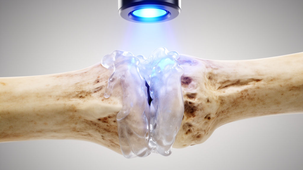
Every year, millions of people suffer complicated bone fractures that require screws, plates, or pins to hold the broken pieces together while the body does the slow work of healing. These metal implants have served orthopedic surgery for decades, but they come with real costs: they are expensive, they sometimes must be surgically removed once the bone has knitted, and they carry a persistent risk of infection. A research team writing in Bioengineering & Translational Medicine now reports a fundamentally different approach — a flowable, injectable bone adhesive that can be painted into an irregular, shattered fracture gap, hardened on demand with 20 seconds of blue-violet light, and then gradually replaced by genuinely new bone as it degrades.
The material, called GelMA–nHA–PEGDA, is built from three components that each play a distinct role. Gelatin methacryloyl, or GelMA, is derived from partially hydrolyzed collagen and retains the cell-interactive motifs and enzyme-cleavable sites of the body’s own structural protein. Polyethylene glycol diacrylate, or PEGDA, is a hydrophilic, biocompatible polymer whose internal architecture resembles the natural extracellular matrix. The third ingredient is nanoscale hydroxyapatite, the calcium phosphate mineral that gives real bone its hardness. Rather than simply mixing mineral powder into a polymer, the team coaxed hydroxyapatite to grow directly onto the GelMA–PEGDA network — a process called biomineralization that imitates how osteoblasts, the bone-forming cells, deposit mineral onto collagen templates during natural osteogenesis.
The chemistry of that mineralization is elegantly simple. Before photocuring, the researchers added calcium nitrate, diammonium hydrogen phosphate, citric acid, and sodium hydroxide directly into the liquid precursor. The negatively charged carboxyl groups on GelMA attract positively charged calcium ions, which in turn neutralize phosphate ions and create nucleation sites on the hydrogel surface. Calcium and phosphate then migrate toward the surface and precipitate as hydroxyapatite, completing the mineralization. Electron microscopy confirmed the result: the original hydrogel’s smooth, porous structure — pores between 10 and 100 micrometers — became denser, with hydroxyapatite nanorods roughly 100 nanometers long and 15 nanometers wide uniformly coating the pore walls. Energy-dispersive X-ray analysis measured a calcium-to-phosphorus ratio of 1.48, closely matching the 1.50 expected for true hydroxyapatite, and X-ray diffraction showed sharp crystalline peaks replacing the amorphous signature of the unmineralized gel.
What makes the material genuinely practical for the operating room is its photosensitivity. The precursor is a warm, molten liquid that can be injected into a fracture gap in a minimally invasive fashion. A patented delivery system then threads an optical fiber through a guiding sleeve to the fracture site and floods it with 405-nanometer light for just 20 seconds. That exposure triggers radical polymerization of the carbon–carbon double bonds in both GelMA and PEGDA, crosslinking the network in place. In tests on fresh chicken femur fragments preconditioned at body temperature, the cured adhesive bonded the two fracture ends strongly enough to support a 500-gram weight in a vertical orientation, and the bond remained stable while submerged in warm saline for 72 hours. The adhesive also stuck to wood, metal, glass, polymers, and other organic surfaces, a versatility that reflects the abundance of hydrogen-bonding groups — hydroxyl, amide, carbonyl, and ether — that grip complementary chemical features on tissue surfaces.
Mechanical testing showed that biomineralization and photocuring work synergistically. In uniaxial compression, the mineralized, cured adhesive tolerated significantly higher maximum pressure before failure than unmineralized material, with elevated modulus and toughness. Three-point bending and lap-shear tests, which probe resistance to vertical and shear stresses respectively, likewise showed substantially higher maximum loads after photocuring. The team reports immediate tensile strength of 0.08 megapascals and lap-shear strength of 0.15 megapascals — figures that exceed representative fibrin-based surgical sealants, though they remain below some experimental polyethylene glycol glues and polymethyl methacrylate bone interfaces. The researchers argue that the combination of rapid application, adequate strength, and regenerative activity could still support standalone use in selected fractures where traditional internal fixation is impractical.
Swelling and degradation behavior matters as much as strength, because an adhesive that lingers too long can block tissue ingrowth. Over 40 days of immersion in body-temperature saline, the material swelled rapidly — more than 50 percent within three days as water penetrated the hydrophilic network — before reaching equilibrium around day 25, when hydrogen bonds among carbonyl groups tightened the crosslinked structure. Degradation proceeded gradually over the same period and accelerated noticeably in the presence of collagenase type II, the enzyme that cleaves the GelMA component. That enzyme-sensitivity is a feature, not a flaw: it means the adhesive should break down in step with the body’s own remodeling machinery, yielding space for new tissue rather than persisting as a permanent barrier the way slow-degrading cyanoacrylate glues can.
Biocompatibility assays with mouse bone marrow-derived mesenchymal stem cells were encouraging. Cells cultured with material extracts showed dose-dependent growth that peaked at 75 percent extract concentration, and adhesion tests at 1, 4, and 7 hours found significantly more cells attached to GelMA–nHA–PEGDA-coated surfaces than to uncoated wells. Live/dead staining revealed predominantly living cells and a higher relative growth rate than either blank controls or plain GelMA. The mineralized adhesive then demonstrated true osteogenic power: Alizarin red staining showed abundant calcium nodules after three weeks of culture, alkaline phosphatase staining — a marker of early osteoblast differentiation — was strongest in the adhesive group, and western blot, quantitative PCR, and immunocytochemistry all confirmed upregulation of the master osteogenic regulators ALP, RUNX2, osteopontin, and collagen 1A1.
The decisive test came in living animals. In a mouse femoral fracture model, 63 animals were randomly assigned to three groups: untreated fractures, conventional Kirschner-wire fixation, and adhesive-only repair with a 20-second light cure. X-ray imaging scored by the Lane–Sandhu system showed that the adhesive group healed significantly better than untreated fractures at both two and four weeks, and by four weeks both the adhesive and pin groups had achieved radiographic union. Histology told an even stronger story: the adhesive group earned the highest callus maturity scores and produced the largest area of new bone, exceeding even the metal-pin group. Three-point bending tests confirmed that adhesive-repaired femurs bore higher maximal loads and stresses than unrepaired bones, and footprint gait analysis showed that mice treated with the adhesive walked with a wide, confident hindlimb stance while untreated and pinned animals dragged their injured limbs.
Micro-computed tomography revealed why the adhesive outperformed the metal pin. Reconstructed three-dimensional images showed complete fracture healing in both treated groups, but the adhesive group displayed significantly greater bone surface, bone volume, trabecular thickness, and trabecular number, with tighter trabecular spacing — a denser, better-organized repair. Immunofluorescent staining pointed to the mechanism: elevated alkaline phosphatase and osteocalcin confirmed active bone formation, while strong signal for paired related homeobox 1, a stem cell marker, showed that the biomineralized gel was actively recruiting bone marrow mesenchymal stem cells to the fracture site. Elevated type II collagen deposition indicated vigorous extracellular matrix assembly. In essence, the adhesive does not merely hold bone still — it behaves like a scaffold of newly forming bone, summoning the cells that build the real thing and switching on their genetic programs as it dissolves.
The implications reach beyond one material. By reproducing the composition and architecture of regenerating bone — collagen-derived polymer, hydroxyapatite mineral, and a degradable, cell-friendly matrix — the team has shown that a fracture fixation device can double as a regenerative medicine product. An all-in-one adhesive that flows into comminuted fracture gaps, cures in seconds under fiber-optic light, bonds to wet tissue, and then hands the job over to the patient’s own stem cells could reduce the burden of implant removal, lower infection risk, and make minimally invasive treatment feasible for injury patterns that currently demand extensive surgery. The reported strengths still fall short of the toughest load-bearing applications, and the work rests on mouse models with a 20-second cure window that will need refinement in larger animals and humans. But as a proof of concept, the study makes a compelling case that the fastest way to fix a broken bone may be to imitate the way bone builds itself.
Subject of Research: A biomineralized photocurable hydrogel bone adhesive for fracture fixation and skeletal regeneration
Article Title: An osteogenesis mimicking biomineralized bone adhesive for rapid skeletal regeneration
Article References: Liu, Y., Luo, H., Guo, B., Chen, X., Stoddart, M. J., Chen, W., Yang, X., Zhu, Y., & Zhu, J. (2026). An osteogenesis mimicking biomineralized bone adhesive for rapid skeletal regeneration. Bioengineering & Translational Medicine, Article e70182. https://doi.org/10.1002/btm2.70182
Image Credits: AI Generated
DOI: 10.1002/btm2.70182
Keywords: bone adhesive, biomineralization, hydroxyapatite, GelMA, PEGDA, photocuring, fracture repair, mesenchymal stem cells, osteogenesis, tissue engineering, regenerative medicine, hydrogel
Cite Scienmag News
Denise Maddox. (October 9, 2026). Light-Cured Bone Glue That Mimics Natural Bone Mineral Speeds Fracture Healing. Scienmag. https://scienmag.com/light-cured-bone-glue-that-mimics-natural-bone-mineral-speeds-fracture-healing/
Denise Maddox. "Light-Cured Bone Glue That Mimics Natural Bone Mineral Speeds Fracture Healing." Scienmag, 9 October 2026, https://scienmag.com/light-cured-bone-glue-that-mimics-natural-bone-mineral-speeds-fracture-healing/. Accessed 9 October 2026.
Denise Maddox. "Light-Cured Bone Glue That Mimics Natural Bone Mineral Speeds Fracture Healing." Scienmag. October 9, 2026. https://scienmag.com/light-cured-bone-glue-that-mimics-natural-bone-mineral-speeds-fracture-healing/


